
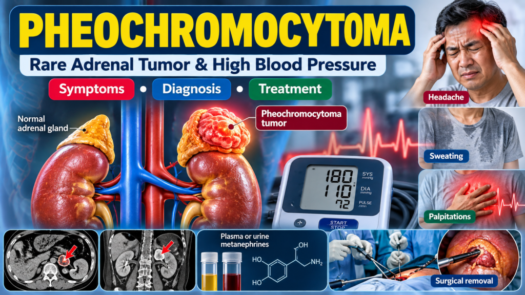
Pheochromocytoma is a rare tumor that develops from chromaffin cells, most commonly in the adrenal glands. These tumors can produce excessive amounts of catecholamines, particularly adrenaline (epinephrine) and noradrenaline (norepinephrine), which can cause episodes of high blood pressure, rapid heartbeat, sweating, headache, and other symptoms.
Although pheochromocytoma is uncommon, it is an important condition to recognize because untreated disease can lead to serious cardiovascular complications.
What Is Pheochromocytoma?
The adrenal glands are small glands located above each kidney. They produce several important hormones, including adrenaline and noradrenaline, which help the body respond to stress.
A pheochromocytoma arises from hormone-producing cells in the adrenal medulla. Most pheochromocytomas are located inside the adrenal glands, although related tumors called paragangliomas can develop outside the adrenal glands along sympathetic or parasympathetic nerves.
These tumors may continuously or intermittently release excessive catecholamines into the bloodstream.
Common Symptoms
Symptoms can vary considerably from person to person. Some patients experience sudden attacks, while others may have persistent symptoms.
Common symptoms include:
- High blood pressure, which may be persistent or occur in episodes
- Severe or pounding headache
- Palpitations or rapid heartbeat
- Excessive sweating
- Tremor or shaking
- Anxiety or a feeling of impending doom
- Pale or flushed appearance
- Chest discomfort
- Shortness of breath
- Dizziness
- Abdominal or back pain
- Weight loss
- Heat intolerance
A classic pattern is an episode of headache, sweating, and palpitations associated with hypertension. However, not every patient has all three symptoms.
Some people may have few or no obvious symptoms, and the tumor may be discovered incidentally during abdominal imaging performed for another reason.
When Should Pheochromocytoma Be Suspected?
Doctors may consider pheochromocytoma in patients with:
- Episodic or difficult-to-control hypertension
- Hypertension at a young age
- Unexplained episodes of palpitations, sweating, and headache
- Hypertensive crises without an obvious cause
- An adrenal mass discovered on CT or MRI
- A previous or family history of pheochromocytoma or paraganglioma
- Certain inherited genetic syndromes associated with pheochromocytoma
Pheochromocytoma should also be considered when symptoms are triggered by certain medications, anesthesia, surgery, or other situations that can provoke catecholamine release.
Causes and Risk Factors
Most pheochromocytomas occur sporadically, but a significant proportion are associated with inherited genetic conditions.
Genetic syndromes associated with pheochromocytoma and paraganglioma include:
- Multiple endocrine neoplasia type 2 (MEN2)
- Von Hippel–Lindau disease
- Neurofibromatosis type 1 (NF1)
- Hereditary paraganglioma syndromes
- SDH gene-related disorders
Because of these associations, genetic evaluation may be appropriate, particularly in younger patients, patients with bilateral or multifocal tumors, those with paraganglioma, or patients with a family history.
How Is Pheochromocytoma Diagnosed?
Diagnosis generally involves biochemical testing followed by imaging.
1. Biochemical Testing
The preferred initial tests are measurements of:
- Plasma free metanephrines, or
- 24-hour urinary fractionated metanephrines
Metanephrines are metabolites of catecholamines and may remain elevated even when catecholamine release from the tumor is intermittent.
Testing conditions are important because stress, certain medications, caffeine, nicotine, acute illness, and improper sampling conditions can sometimes produce abnormal results.
A markedly elevated result is more concerning for pheochromocytoma than a mild elevation, which may require careful clinical interpretation and sometimes repeat testing.
2. Imaging
Once biochemical testing strongly suggests pheochromocytoma, imaging is performed to identify the tumor.
Common imaging studies include:
- CT scan of the abdomen and pelvis
- MRI, particularly when radiation exposure should be minimized or when evaluating certain extra-adrenal tumors
Additional functional imaging may be considered when disease is multifocal, metastatic, recurrent, or difficult to localize.
Treatment
The main treatment for a localized pheochromocytoma is generally surgical removal of the tumor.
However, surgery must be carefully prepared because manipulation of the tumor can cause a sudden release of catecholamines, resulting in severe hypertension, arrhythmias, or cardiovascular complications.
Preoperative Medical Preparation
Patients are usually treated with alpha-adrenergic blockade before surgery.
Commonly used medications include:
- Phenoxybenzamine
- Selective alpha-1 blockers such as doxazosin
Adequate fluid and salt intake may also be encouraged when appropriate to restore the patient’s intravascular volume.
Important: Alpha Blockade Before Beta Blockade
If a patient requires beta-blockade for significant tachycardia or arrhythmia, alpha blockade should be established first.
Giving a beta-blocker before adequate alpha blockade can result in unopposed alpha-adrenergic stimulation and potentially severe hypertension.
This is an important safety principle in the management of pheochromocytoma.
Surgery
Surgical options depend on the location, size, number of tumors, genetic background, and whether the disease has spread.
For many localized adrenal tumors, laparoscopic or minimally invasive adrenalectomy can be performed.
More extensive surgery may be required for large, invasive, multifocal, or extra-adrenal tumors.
Patients require careful blood pressure and cardiovascular monitoring before, during, and after surgery.
What Happens After Surgery?
After successful removal of a catecholamine-producing tumor, blood pressure and catecholamine levels often improve substantially.
However, long-term follow-up is important because pheochromocytoma can recur, particularly in patients with hereditary disease or certain tumor characteristics.
Follow-up may include:
- Blood pressure monitoring
- Plasma or urinary metanephrine testing
- Periodic imaging when clinically indicated
- Genetic evaluation when appropriate
Can Pheochromocytoma Be Malignant?
Most pheochromocytomas are not immediately classified as malignant based only on their microscopic appearance.
The clinically important definition of malignancy is the presence of metastases to sites where chromaffin tissue normally does not occur, such as lymph nodes, bone, liver, or lungs.
Metastatic disease requires specialized management and may involve surgery, radiotherapy, radionuclide therapy, systemic therapy, or other treatments depending on the individual case.
Pheochromocytoma and Pregnancy
Pheochromocytoma during pregnancy is rare but potentially serious for both the mother and fetus.
Unexplained severe or episodic hypertension during pregnancy, particularly when accompanied by headache, palpitations, or sweating, requires careful evaluation.
Management should involve a multidisciplinary team with expertise in endocrinology, maternal-fetal medicine, anesthesia, and surgery.
Key Takeaways
Pheochromocytoma is a rare but important cause of secondary hypertension.
Remember these key points:
- Consider pheochromocytoma in patients with episodic hypertension, headache, sweating, and palpitations.
- Plasma free metanephrines or urinary fractionated metanephrines are important initial biochemical tests.
- CT or MRI is generally used to localize the tumor after biochemical evaluation.
- Surgery is the main treatment for localized disease.
- Adequate alpha-adrenergic blockade before surgery is essential.
- If beta-blockade is needed, establish adequate alpha blockade first.
- Consider hereditary disease and genetic testing when clinically appropriate.
- Long-term biochemical and clinical follow-up is important because recurrence can occur.
When Should You See a Doctor?
If you experience repeated episodes of severe headache, sweating, palpitations, tremor, or markedly elevated blood pressure, particularly when the episodes occur suddenly and without an obvious trigger, medical evaluation is recommended.
An adrenal mass discovered incidentally on CT or MRI should also be appropriately evaluated to determine whether it produces hormones.
Early recognition and appropriate treatment can significantly reduce the risk of serious cardiovascular complications.
Medical information on this page is for educational purposes and does not replace an individualized medical consultation.
FAQ Section
1. What is pheochromocytoma?
Pheochromocytoma is a rare tumor arising from catecholamine-producing cells, usually in the adrenal gland. It can produce excessive adrenaline and noradrenaline, resulting in high blood pressure and other symptoms.
2. What are the most common symptoms of pheochromocytoma?
Common symptoms include episodic or persistent high blood pressure, severe headache, sweating, palpitations, tremor, anxiety, chest discomfort and sometimes weight loss.
3. What is the classic triad of pheochromocytoma?
The classic symptom combination is headache, sweating, and palpitations, often associated with hypertension. However, many patients do not have all three symptoms.
4. How is pheochromocytoma diagnosed?
Initial biochemical evaluation usually involves plasma free metanephrines or urinary fractionated metanephrines. CT or MRI may then be used to locate the tumor.
5. Can pheochromocytoma cause high blood pressure?
Yes. Excess catecholamine production can cause severe, episodic, or persistent hypertension and is an important cause of secondary hypertension.
6. Is pheochromocytoma curable?
Localized pheochromocytoma can often be treated successfully with surgical removal. Long-term follow-up is important because recurrence can occur.
7. What treatment is given before pheochromocytoma surgery?
Patients generally receive alpha-adrenergic blockade before surgery to control blood pressure and reduce the risk of a dangerous catecholamine surge during tumor removal.
8. Can pheochromocytoma be hereditary?
Yes. Pheochromocytoma can occur as part of inherited conditions such as MEN2, von Hippel–Lindau disease, NF1, and hereditary paraganglioma syndromes.
9. Can pheochromocytoma be malignant?
Some pheochromocytomas can metastasize. Malignancy is established by the presence of metastases to sites where chromaffin tissue normally does not exist.
10. When should I see a doctor?
Medical evaluation is appropriate if you have recurrent episodes of severe headache, sweating, palpitations, unexplained hypertension, or an adrenal mass discovered on imaging.