
Introduction
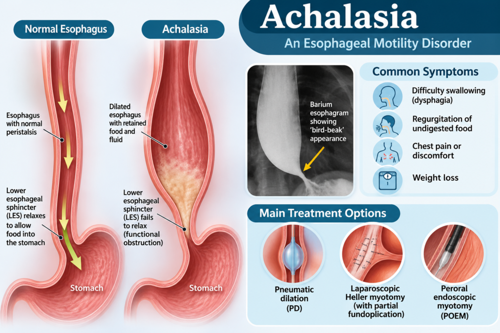
Achalasia is a rare but important esophageal motility disorder characterized by impaired relaxation of the lower esophageal sphincter (LES) and loss of normal esophageal peristalsis. These abnormalities prevent food and liquids from passing normally from the esophagus into the stomach.
Patients commonly experience progressive difficulty swallowing (dysphagia), regurgitation of undigested food, chest discomfort, and weight loss. Without appropriate treatment, achalasia can lead to significant esophageal dilation, nutritional deficiencies, aspiration, and impaired quality of life.
Although there is currently no treatment that reverses the underlying neuronal damage, several effective therapeutic options can relieve symptoms and improve esophageal emptying.
This article reviews the causes, clinical manifestations, diagnostic approach, complications, and evidence-based treatment strategies for achalasia, based on current clinical guidelines and evidence available in 2026.
1. What Is Achalasia?
Achalasia is a primary esophageal motor disorder involving two major abnormalities:
- Failure of the lower esophageal sphincter to relax adequately during swallowing.
- Absence of normal peristaltic contractions in the esophageal body.
Normally, swallowing triggers coordinated muscular contractions that propel food toward the stomach while the LES relaxes to allow food to pass.
In achalasia, this coordinated process is disrupted. Food and liquids accumulate in the esophagus, resulting in progressive dilation and symptoms of dysphagia and regurgitation.
Achalasia is uncommon, with an estimated annual incidence of approximately 1–2 cases per 100,000 people.
2. Causes and Pathophysiology of Achalasia
Primary achalasia
The exact cause of primary achalasia remains incompletely understood.
The principal pathological mechanism is progressive degeneration of inhibitory neurons within the myenteric (Auerbach’s) plexus of the esophageal wall.
These neurons normally release neurotransmitters, particularly:
- Nitric oxide (NO).
- Vasoactive intestinal peptide (VIP).
These neurotransmitters are essential for LES relaxation during swallowing.
When inhibitory neurons are lost or dysfunctional, the following changes occur:
- Impaired relaxation of the LES.
- Increased or inadequately reduced LES pressure.
- Loss of normal esophageal peristalsis.
- Progressive retention of food and liquids.
- Esophageal dilation and, in advanced cases, a sigmoid-shaped esophagus.
Autoimmune mechanisms and genetic susceptibility have been proposed as contributing factors. Some studies also suggest a possible association with viral triggers, although a definitive causal relationship has not been established.
Secondary achalasia (pseudoachalasia)
Pseudoachalasia describes a condition that resembles primary achalasia clinically and on manometry but results from another underlying disease.
Important causes include:
- Gastroesophageal junction or gastric cardia malignancy.
- Esophageal cancer.
- External compression or infiltrative malignancy.
- Chagas disease caused by Trypanosoma cruzi.
- Other infiltrative or postsurgical conditions.
Chagas disease is particularly important in patients with relevant epidemiological exposure, especially in endemic regions of Latin America.
Clinical warning: Always consider pseudoachalasia in older patients with rapidly progressive dysphagia, substantial weight loss, or a short symptom history.
3. Clinical Manifestations of Achalasia
Symptoms generally develop gradually and progress over months or years.
Common symptoms
1. Dysphagia
- Difficulty swallowing both solid foods and liquids.
- Often progressive.
- Patients may need to drink water to help food pass.
- Food may feel stuck behind the sternum.
2. Regurgitation
- Return of undigested food or saliva into the mouth.
- Frequently occurs after meals or when lying down.
- May cause nocturnal coughing or choking.
3. Chest pain
- Retrosternal discomfort or pressure.
- May occur during or after meals.
- Can resemble cardiac chest pain.
4. Weight loss
- Results from reduced oral intake and difficulty swallowing.
- May become significant in advanced disease.
5. Respiratory symptoms
Retention of food and secretions can result in aspiration, causing:
- Chronic cough.
- Recurrent respiratory infections.
- Nocturnal choking.
- Aspiration pneumonia.
Other possible manifestations
- Heartburn-like symptoms.
- Excessive salivation.
- Halitosis.
- Nutritional deficiencies.
- Sleep disturbance.
Heartburn does not necessarily indicate GERD. Retained food and fermentation within the esophagus can produce symptoms that resemble acid reflux.
4. Classification of Achalasia
High-resolution esophageal manometry (HRM) classifies achalasia into three major subtypes according to the Chicago Classification version 4.0.
Type I: Classic achalasia
- Elevated integrated relaxation pressure (IRP).
- Absent normal peristalsis.
- No significant panesophageal pressurization.
This subtype is associated with poor esophageal contractility.
Type II: Achalasia with panesophageal pressurization
- Elevated IRP.
- Absent normal peristalsis.
- Panesophageal pressurization in at least 20% of swallows.
This subtype generally has the most favorable treatment response among the three types.
Type III: Spastic achalasia
- Elevated IRP.
- Absent normal peristalsis.
- Premature or spastic contractions in at least 20% of swallows.
This subtype is associated with spastic esophageal contractions and may present with prominent chest pain.
Clinical importance: Achalasia subtype helps guide treatment selection. POEM is particularly useful in type III disease because it allows a longer, tailored myotomy.
5. Diagnosis of Achalasia
The diagnosis should combine clinical presentation, upper gastrointestinal endoscopy, esophageal manometry, and radiological assessment.
A. Upper gastrointestinal endoscopy (EGD)
Upper endoscopy is recommended in patients with suspected achalasia.
Typical findings include:
- Retained food and saliva in the esophagus.
- Dilated esophageal lumen.
- Tight gastroesophageal junction.
- Resistance when passing the endoscope through the LES.
Endoscopy is particularly important to exclude mechanical obstruction, malignancy, and pseudoachalasia.
A normal-looking endoscopy does not exclude achalasia.
B. High-resolution esophageal manometry (HRM)
HRM is the gold-standard investigation for confirming achalasia.
Typical findings include:
- Elevated median integrated relaxation pressure (IRP).
- Absent normal esophageal peristalsis.
- Abnormal pressurization or spastic contractions depending on the subtype.
The diagnostic IRP threshold depends on the manometry system, catheter position, and laboratory reference values. An isolated elevated IRP should not be interpreted without the complete manometric pattern and clinical context.
HRM also identifies the three major achalasia subtypes.
C. Timed barium esophagram (TBE)
A timed barium esophagram is a useful investigation for diagnosing achalasia and evaluating esophageal emptying.
Typical findings include:
- Dilated esophagus.
- Smooth narrowing at the gastroesophageal junction.
- Characteristic bird-beak appearance.
- Retained barium with delayed esophageal emptying.
A standardized timed study commonly measures the height of the barium column at 1, 2, and 5 minutes after ingestion.
TBE is also valuable for assessing treatment response and suspected recurrence.
D. Functional lumen imaging probe (FLIP)
FLIP is an adjunctive diagnostic technique that evaluates esophageal distensibility and gastroesophageal junction opening.
It can be particularly helpful when:
- HRM results are inconclusive.
- The diagnosis remains uncertain.
- Additional physiological assessment is required.
FLIP should complement rather than routinely replace HRM.
E. CT scan or endoscopic ultrasound
CT imaging or endoscopic ultrasound may be indicated when pseudoachalasia is suspected, particularly in patients with:
- Older age at symptom onset.
- Rapidly progressive dysphagia.
- Marked or unexplained weight loss.
- Short symptom duration.
- Difficult passage of the endoscope through the gastroesophageal junction.
These investigations help identify an underlying malignancy or extrinsic compression.
6. Differential Diagnosis
Conditions that may resemble achalasia include:
- Gastroesophageal reflux disease (GERD).
- Esophageal cancer.
- Esophageal stricture.
- Eosinophilic esophagitis.
- Esophageal spasm.
- Esophagogastric junction outflow obstruction (EGJOO).
- Systemic sclerosis-associated esophageal dysmotility.
- Chagas disease.
- Pseudoachalasia.
A careful clinical assessment and appropriate investigations are necessary to distinguish these conditions.
7. Treatment of Achalasia
The main treatment objective is to reduce resistance at the LES and improve esophageal emptying.
Treatment does not restore the damaged enteric neurons or normal peristalsis.
Treatment selection should consider:
- Achalasia subtype.
- Patient age and comorbidities.
- Esophageal anatomy.
- Previous treatment.
- Patient preference.
- Local availability and procedural expertise.
A. Pneumatic dilation (PD)
Pneumatic dilation is an endoscopic treatment involving the use of a balloon to disrupt the LES muscle fibers.
The procedure helps reduce LES pressure and improve the passage of food into the stomach.
Advantages:
- Effective for type I and type II achalasia.
- Less invasive than surgical myotomy.
- Can be repeated if symptoms recur.
Limitations and complications:
- Esophageal perforation.
- Gastroesophageal reflux.
- Chest discomfort.
- Recurrence of symptoms.
Graded pneumatic dilation commonly begins with a 3.0-cm balloon, with subsequent dilation depending on clinical response and patient characteristics.
Younger male patients may have a higher risk of treatment failure and may require an individualized initial strategy.
B. Laparoscopic Heller myotomy (LHM)
Heller myotomy is a surgical procedure in which the muscle fibers of the LES are divided to facilitate esophageal emptying.
It is commonly combined with a partial fundoplication to reduce postoperative gastroesophageal reflux.
Common fundoplication techniques include:
- Dor fundoplication (anterior partial wrap).
- Toupet fundoplication (posterior partial wrap).
Advantages:
- Durable symptom improvement.
- Effective for type I and type II achalasia.
- Allows simultaneous partial fundoplication to reduce reflux.
Potential complications:
- Gastroesophageal reflux.
- Mucosal perforation.
- Bleeding.
- Postoperative dysphagia.
- Recurrence of symptoms.
C. Peroral endoscopic myotomy (POEM)
POEM is a minimally invasive endoscopic procedure in which the endoscopist creates a submucosal tunnel and divides the circular muscle fibers of the esophagus and LES.
It does not require external surgical incisions.
POEM has become an important treatment option because of its high clinical effectiveness and ability to perform an extended myotomy.
Advantages:
- High clinical success rates.
- Particularly useful in type III achalasia.
- Allows a longer myotomy tailored to the extent of spastic contractions.
- Avoids external surgical incisions.
Potential complications:
- Gastroesophageal reflux.
- Mucosal injury.
- Bleeding.
- Pneumoperitoneum or pneumomediastinum.
- Rare serious procedural complications.
One important limitation of POEM is the increased risk of postprocedural GERD compared with treatments that include an antireflux procedure.
Patients should be counseled about reflux symptoms, objective reflux assessment, and appropriate proton pump inhibitor (PPI) therapy.
D. Botulinum toxin injection
Botulinum toxin is injected endoscopically into the LES to reduce sphincter tone.
It inhibits acetylcholine release at neuromuscular junctions, temporarily relaxing the sphincter.
Indications:
- Patients who are not suitable candidates for pneumatic dilation, POEM, or surgery.
- Frail elderly patients.
- Patients with significant comorbidities or high procedural risk.
Limitations:
- Temporary symptom improvement.
- Frequent recurrence.
- Repeated injections may be necessary.
- Not recommended as definitive first-line treatment in patients who are suitable for definitive therapy.
E. Pharmacological treatment
Medications have a limited role in achalasia because they do not provide durable control.
Options include:
- Nitrates.
- Calcium channel blockers, such as nifedipine.
These medications may temporarily reduce LES pressure but have limited efficacy and potential adverse effects, including headache, dizziness, and hypotension.
They are generally reserved for patients who cannot undergo definitive treatment.
8. Choosing the Best Treatment According to Achalasia Subtype
| Achalasia subtype | Preferred treatment options |
|---|---|
| Type I | POEM, pneumatic dilation, or Heller myotomy with fundoplication |
| Type II | POEM, pneumatic dilation, or Heller myotomy with fundoplication |
| Type III | POEM is generally preferred; tailored Heller myotomy may be considered |
What do the latest guidelines recommend?
The 2024 update to the SAGES guidelines, published in 2025, provides important recommendations:
- POEM is conditionally suggested over pneumatic dilation for adult achalasia.
- POEM with appropriate PPI use or Heller myotomy with fundoplication are both reasonable options for type I and type II achalasia.
- POEM is favored over Heller myotomy with fundoplication for type III achalasia.
The American Gastroenterological Association (AGA) 2024 Clinical Practice Update also emphasizes comprehensive diagnostic evaluation and individualized treatment selection.
The choice between treatment modalities should be made through shared decision-making, considering the patient’s condition, preferences, and availability of experienced specialists.
9. Management After Treatment
Successful treatment does not eliminate the need for follow-up.
Patients should be monitored for:
- Persistent or recurrent dysphagia.
- Regurgitation.
- Gastroesophageal reflux.
- Esophagitis.
- Esophageal emptying.
- Nutritional status.
Post-treatment assessment
The Eckardt score can help evaluate clinical response.
It assesses four symptoms:
- Dysphagia.
- Regurgitation.
- Chest pain.
- Weight loss.
Each item is scored from 0 to 3, producing a total score of 0–12.
A score of 3 or less is commonly used as a marker of clinical success, although symptom assessment alone does not always reflect adequate esophageal emptying.
Timed barium esophagram is useful when symptoms persist or recur.
Endoscopy should be considered when reflux-related complications, esophagitis, or recurrent obstruction are suspected.
Patients treated with POEM should receive appropriate counseling and follow-up for postprocedural GERD. PPI therapy and objective reflux assessment should be individualized.
10. Complications of Achalasia
Untreated or advanced achalasia can lead to several complications.
1. Esophageal dilation
Chronic food retention and impaired emptying result in progressive esophageal enlargement.
2. Megaesophagus and sigmoid esophagus
Advanced disease may produce marked dilation and tortuosity, making treatment more challenging.
3. Aspiration pneumonia
Retained food and secretions can enter the respiratory tract, particularly during sleep.
4. Malnutrition and weight loss
Progressive dysphagia may cause inadequate nutritional intake.
5. Esophagitis
Prolonged retention of food and fermentation products can cause mucosal inflammation.
6. Esophageal cancer
Achalasia is associated with an increased relative risk of esophageal cancer, particularly squamous cell carcinoma.
However, the absolute risk remains relatively low, and routine endoscopic cancer surveillance at fixed intervals is not universally recommended by current guidelines.
New or changing symptoms should prompt appropriate investigation.
11. Lifestyle and Dietary Recommendations
Lifestyle modification can help reduce symptoms, although it cannot correct the underlying motility disorder.
Patients may benefit from:
- Eating smaller, more frequent meals.
- Chewing food thoroughly.
- Eating slowly.
- Drinking water with meals if tolerated.
- Avoiding foods that are difficult to swallow.
- Remaining upright after meals.
- Avoiding meals immediately before bedtime.
- Elevating the head of the bed when nocturnal regurgitation occurs.
- Maintaining adequate hydration and nutrition.
Patients with significant dysphagia or weight loss should receive nutritional assessment and individualized dietary advice.
12. Prognosis
Achalasia is a chronic condition, but most patients can achieve substantial symptom improvement with appropriate treatment.
POEM, pneumatic dilation, and Heller myotomy are effective treatment options, although recurrence may occur.
Type II achalasia generally has a favorable response to treatment, while type III disease may require a longer myotomy.
Long-term follow-up is important because patients can develop recurrent obstruction, GERD, esophagitis, or progressive esophageal dilation.
Conclusion
Achalasia is a rare esophageal motility disorder characterized by impaired LES relaxation and absent normal esophageal peristalsis.
The diagnosis is based primarily on high-resolution esophageal manometry, supported by upper gastrointestinal endoscopy and timed barium esophagram.
Treatment options include pneumatic dilation, laparoscopic Heller myotomy with partial fundoplication, and peroral endoscopic myotomy (POEM).
Current evidence supports individualized treatment selection. POEM is particularly valuable in type III achalasia, while POEM, pneumatic dilation, and Heller myotomy remain effective options for type I and type II disease.
Early diagnosis, exclusion of pseudoachalasia, appropriate treatment, and long-term follow-up are essential to improving patient outcomes.
Frequently Asked Questions (FAQ)
1. What is the main cause of achalasia?
Achalasia is primarily caused by degeneration of inhibitory neurons in the esophageal myenteric plexus. This results in impaired LES relaxation and loss of normal esophageal peristalsis. The exact initiating cause remains uncertain.
2. What are the earliest symptoms of achalasia?
The most common early symptom is difficulty swallowing both solid foods and liquids. Patients may also experience regurgitation, chest discomfort, and unexplained weight loss.
3. Is achalasia the same as GERD?
No. GERD results from reflux of gastric contents into the esophagus, whereas achalasia is a motility disorder involving impaired LES relaxation and absent normal peristalsis. However, both conditions can cause heartburn-like symptoms.
4. What is the best test to diagnose achalasia?
High-resolution esophageal manometry is the gold-standard diagnostic test. Upper gastrointestinal endoscopy is also essential to exclude malignancy and other mechanical causes of obstruction.
5. What is the bird-beak sign?
The bird-beak sign is a characteristic radiological finding on a barium esophagram. It describes a dilated esophagus with smooth, tapered narrowing at the gastroesophageal junction.
6. What are the three types of achalasia?
According to Chicago Classification version 4.0:
- Type I: Classic achalasia.
- Type II: Achalasia with panesophageal pressurization.
- Type III: Spastic achalasia.
7. What is the best treatment for achalasia?
There is no single best treatment for every patient. POEM, pneumatic dilation, and laparoscopic Heller myotomy with fundoplication are effective options. POEM is generally preferred for type III achalasia.
8. Is POEM better than Heller myotomy?
Both are effective. POEM is particularly advantageous for type III achalasia because it allows an extended myotomy. However, POEM carries a higher risk of postprocedural GERD. Heller myotomy combined with partial fundoplication offers an antireflux component.
9. Can achalasia be cured completely?
Current treatments improve LES opening and esophageal emptying but do not reverse the underlying neuronal damage. Many patients achieve long-term symptom relief, although recurrence and complications can occur.
10. Can achalasia cause weight loss?
Yes. Difficulty swallowing reduces food intake and can lead to progressive weight loss and malnutrition.
11. Does achalasia increase the risk of esophageal cancer?
Yes. Long-standing achalasia is associated with an increased risk of esophageal cancer, particularly squamous cell carcinoma. Nevertheless, routine endoscopic surveillance at fixed intervals is not currently recommended for every patient.
12. Can medications treat achalasia?
Nitrates and calcium channel blockers may temporarily reduce LES pressure, but their efficacy is limited. They are generally reserved for patients who cannot undergo definitive treatment.
13. What is pseudoachalasia?
Pseudoachalasia is a condition that mimics primary achalasia but is caused by another disease, particularly malignancy at the gastroesophageal junction or gastric cardia. It should be suspected in older patients with rapid symptom progression and substantial weight loss.
14. Can achalasia cause coughing at night?
Yes. Regurgitation of retained food and saliva during sleep can cause nocturnal coughing, choking, and aspiration pneumonia.
15. Does achalasia require lifelong follow-up?
Yes. Long-term follow-up is important to detect symptom recurrence, reflux complications, impaired esophageal emptying, and nutritional problems.
References
- Vaezi MF, Pandolfino JE, Yadlapati RH, Greer KB, Kavitt RT. ACG Clinical Guidelines: Diagnosis and Management of Achalasia. The American Journal of Gastroenterology. 2020;115(9):1393–1411. doi:10.14309/ajg.0000000000000731.
- Calabrese EC, et al. 2024 Update to SAGES Guidelines for the Use of Peroral Endoscopic Myotomy (POEM) in the Treatment of Achalasia. Surgical Endoscopy. Published 2025.
- Yang D, Bechara R, Dunst CM, Konda VJA. Advances in Per-Oral Endoscopic Myotomy. American Gastroenterological Association Clinical Practice Update. Gastroenterology. 2024.
- ASGE Standards of Practice Committee. ASGE Guideline on the Management of Achalasia. Gastrointestinal Endoscopy. 2020;91(2):213–227.
- Yadlapati R, et al. Esophageal Motility Disorders on High-Resolution Manometry: Chicago Classification Version 4.0. Neurogastroenterology & Motility. 2021;33(1). doi:10.1111/nmo.14058.
- Bredenoord AJ, et al. European Guideline on Achalasia – UEG and ESNM Recommendations. United European Gastroenterology Journal. 2020;8(1):13–34.
- Multi-Society Clinical Practice Guideline for the Treatment of Adult Patients With Achalasia. Diseases of the Esophagus. 2026;39(Supplement 1). doi:10.1093/dote/doag077.063.
Medical disclaimer: This article is intended for educational purposes and does not replace individualized medical evaluation, diagnosis, or treatment by a qualified healthcare professional.