
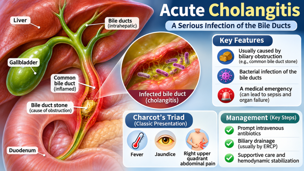
Early recognition, appropriate intravenous antibiotics, supportive care, and timely biliary drainage are essential to improving patient outcomes.
This article reviews the causes, clinical presentation, diagnostic criteria, severity assessment, and evidence-based management of acute cholangitis, incorporating the Tokyo Guidelines (TG18), American Society for Gastrointestinal Endoscopy (ASGE) recommendations, and relevant evidence available through 2026.
1. What is acute cholangitis?
Acute cholangitis is an infection and inflammation of the bile ducts, most commonly resulting from bacterial infection in the presence of biliary obstruction.
Normally, bile flows from the liver through the intrahepatic and extrahepatic bile ducts into the duodenum. When bile flow becomes obstructed, intrabiliary pressure increases, allowing bacteria to proliferate and potentially enter the bloodstream.
The most common cause is choledocholithiasis (common bile duct stones).
Other causes include:
-
Benign or malignant biliary strictures.
-
Pancreatic cancer.
-
Cholangiocarcinoma.
-
Ampullary tumors.
-
Biliary stent obstruction or infection.
-
Post-ERCP complications.
-
Primary sclerosing cholangitis with biliary obstruction.
2. Causes and risk factors
Common causes
1. Common bile duct stones (choledocholithiasis)
This is the leading cause of acute cholangitis. Gallstones migrate from the gallbladder into the common bile duct, causing obstruction and subsequent bacterial infection.
2. Malignant biliary obstruction
Tumors can obstruct bile flow, including:
-
Pancreatic head cancer.
-
Cholangiocarcinoma.
-
Ampullary carcinoma.
-
Gallbladder cancer.
3. Benign biliary obstruction
Possible causes include postoperative biliary strictures, chronic pancreatitis, and inflammatory biliary disease.
4. Biliary instrumentation
ERCP, biliary stent placement, and other biliary procedures can introduce bacteria or lead to subsequent obstruction.
Risk factors
-
History of gallstones.
-
Previous biliary surgery or ERCP.
-
Biliary stent placement.
-
Pancreatic or biliary malignancy.
-
Advanced age.
-
Immunosuppression.
-
Previous episodes of cholangitis.
3. Pathophysiology
The development of acute cholangitis generally involves two important mechanisms:
-
Biliary obstruction: Increased intraductal pressure results from impaired bile drainage.
-
Bacterial infection: Bacteria ascend from the duodenum or enter through instrumentation, proliferate in stagnant bile, and may spread into the bloodstream.
Increased biliary pressure facilitates bacterial and endotoxin translocation into the systemic circulation.
This can lead to:
-
Systemic inflammatory response.
-
Bacteremia.
-
Sepsis.
-
Septic shock.
-
Acute kidney injury and multiorgan dysfunction.
Common causative organisms include:
-
Escherichia coli
-
Klebsiella species
-
Enterococcus species
-
Enterobacter species
-
Pseudomonas aeruginosa (particularly in healthcare-associated infections)
Polymicrobial infections can occur.
4. Clinical manifestations and symptoms
The classical clinical presentation is Charcot’s triad:

-
Fever, often with chills or rigors.
-
Jaundice.
-
Right upper quadrant abdominal pain.
However, Charcot’s triad is not sufficiently sensitive to exclude acute cholangitis when absent.
Other symptoms and signs include:
-
Nausea and vomiting.
-
Malaise and weakness.
-
Dark urine.
-
Pale stools.
-
Pruritus.
-
Hypotension.
-
Confusion or altered mental status.
Reynolds’ pentad
Reynolds’ pentad consists of Charcot’s triad plus:
-
Hypotension.
-
Altered mental status.
This presentation suggests severe cholangitis with sepsis and potentially septic shock.
Patients with suspected cholangitis and hypotension, confusion, or other evidence of organ dysfunction require immediate hospital assessment and urgent treatment.
5. Diagnosis of acute cholangitis
Diagnosis is based on clinical findings, laboratory investigations, and imaging.
The Tokyo Guidelines 2018 (TG18) provide widely used diagnostic criteria.
A. Systemic inflammation
At least one of the following:
-
Fever above 38°C or shaking chills.
-
White blood cell count below 4,000/mm³ or above 10,000/mm³.
-
C-reactive protein (CRP) ≥1 mg/dL.
B. Cholestasis
At least one of the following:
-
Total bilirubin ≥2 mg/dL.
-
ALP, GGT, AST, or ALT greater than 1.5 times the upper limit of normal.
C. Imaging findings
At least one of the following:
-
Biliary duct dilatation.
-
Evidence of the cause of obstruction, such as a common bile duct stone, biliary stricture, or obstructed stent.
Diagnostic classification:
| Classification | Criteria |
|---|---|
| Suspected cholangitis | A + B, or A + C |
| Definite cholangitis | A + B + C |
Importantly, clinicians should not delay initial treatment while waiting for all diagnostic criteria to be fulfilled.
JSHBPS+1
Laboratory investigations
Recommended investigations include:
-
Complete blood count (CBC).
-
CRP.
-
Liver function tests (bilirubin, ALP, GGT, AST, ALT).
-
Renal function and electrolytes.
-
Blood glucose.
-
Coagulation profile (PT/INR).
-
Serum lactate when sepsis or tissue hypoperfusion is suspected.
-
Blood cultures before antibiotics, if this does not delay treatment.
-
Blood cultures and bile cultures where clinically appropriate.
A cholestatic pattern of liver enzyme abnormalities is common, although AST and ALT may also become markedly elevated in acute biliary obstruction.
Imaging investigations
1. Abdominal ultrasound
Usually the first-line imaging investigation.
It can identify:
-
Dilated intrahepatic or extrahepatic bile ducts.
-
Gallstones.
-
Gallbladder abnormalities.
-
Some common bile duct stones.
However, a normal ultrasound does not exclude choledocholithiasis or acute cholangitis.
2. Magnetic resonance cholangiopancreatography (MRCP)
MRCP is a noninvasive investigation that provides detailed visualization of the biliary and pancreatic ducts.
It is useful for identifying:
-
Common bile duct stones.
-
Biliary strictures.
-
Biliary dilatation.
-
Suspected malignant obstruction.
3. Endoscopic ultrasound (EUS)
EUS is particularly useful for detecting small common bile duct stones that may not be visible on ultrasound or MRCP.
4. Computed tomography (CT)
CT is useful when malignancy, pancreatic disease, complications, or alternative abdominal diagnoses are suspected.
5. ERCP
ERCP is primarily a therapeutic procedure in acute cholangitis. It allows biliary decompression, stone extraction, and stent placement.
6. Severity grading according to Tokyo Guidelines (TG18)
Severity assessment is essential because it determines the urgency of biliary drainage and the level of supportive care required.
TG18 classifies acute cholangitis into three grades.
Grade I – Mild cholangitis
Does not meet the criteria for Grade II or Grade III.
-
No organ dysfunction.
-
Usually responds to initial medical treatment, including antibiotics and supportive care.
-
Biliary drainage is considered if the patient does not improve.
Grade II – Moderate cholangitis
At least TWO of the following criteria:
-
WBC count above 12,000/mm³ or below 4,000/mm³.
-
Fever ≥39°C.
-
Age ≥75 years.
-
Total bilirubin ≥5 mg/dL.
-
Albumin below 70% of the lower limit of normal.
These patients generally require early biliary drainage.
Grade III – Severe cholangitis
Defined by dysfunction of at least ONE organ system:
| Organ system | Criteria |
|---|---|
| Cardiovascular | Hypotension requiring dopamine ≥5 μg/kg/min or any dose of norepinephrine |
| Neurological | Disturbance of consciousness |
| Respiratory | PaO₂/FiO₂ ratio below 300 |
| Renal | Oliguria or creatinine above 2 mg/dL |
| Hepatic | PT-INR above 1.5 |
| Hematological | Platelet count below 100,000/mm³ |
These patients require urgent resuscitation, intensive monitoring, and emergency biliary decompression as soon as feasible.
Wiley Online Library+1
7. Management of acute cholangitis
The treatment of acute cholangitis involves three major components:
-
Hemodynamic stabilization and supportive care.
-
Early appropriate intravenous antibiotics.
-
Relief of biliary obstruction through drainage.
Antibiotics alone are not sufficient in most patients with persistent biliary obstruction. Source control is essential.
A. Initial stabilization
Assess airway, breathing, circulation, mental status, and vital signs.
Initial management includes:
-
Establish intravenous access.
-
Monitor blood pressure, heart rate, oxygen saturation, and urine output.
-
Administer IV crystalloid fluids for hypovolemia or sepsis-associated hypoperfusion, with reassessment to avoid fluid overload.
-
Provide oxygen if hypoxemia is present.
-
Obtain blood cultures before antibiotics when feasible without delaying treatment.
-
Monitor renal function, electrolytes, lactate, and coagulation.
-
Initiate vasopressors, preferably norepinephrine, if hypotension persists despite appropriate fluid resuscitation.
Patients with septic shock or significant organ dysfunction should be managed in an intensive care setting when available.
B. Empirical antibiotic therapy
Antibiotics should be initiated promptly after obtaining appropriate cultures, without delaying treatment in critically ill patients.
Empirical treatment should cover common enteric Gram-negative organisms and, when indicated, anaerobes and Enterococcus.
The choice depends on:
-
Severity of infection.
-
Community-acquired versus healthcare-associated infection.
-
Previous antibiotic exposure.
-
History of biliary instrumentation or stent placement.
-
Local antimicrobial resistance patterns.
-
Renal and hepatic function.
Examples of empirical IV antibiotic regimens in adults:
| Antibiotic | Typical adult regimen |
|---|---|
| Ceftriaxone | 2 g IV every 24 hours |
| Ceftriaxone + metronidazole | 2 g IV every 24 hours + 500 mg IV every 8–12 hours |
| Piperacillin/tazobactam | 4.5 g IV every 6–8 hours, depending on infusion protocol |
| Cefepime + metronidazole | Cefepime 2 g IV every 8–12 hours + metronidazole 500 mg IV every 8–12 hours |
| Meropenem | 1 g IV every 8 hours |
These are example regimens, not universal prescriptions. Adjust for renal function, infection severity, local susceptibility data, and institutional protocols.
Important considerations:
-
Ceftriaxone plus metronidazole is a possible option for selected community-acquired infections.
-
Piperacillin/tazobactam is often appropriate when broader Gram-negative and anaerobic coverage is needed.
-
Carbapenems may be appropriate for severe healthcare-associated infections or suspected ESBL-producing organisms, depending on local resistance patterns.
-
Consider additional Enterococcus coverage in selected healthcare-associated infections, particularly in patients with previous biliary instrumentation or transplantation.
-
Antibiotic therapy should be narrowed according to blood and bile culture results.
Duration of antibiotics
Following adequate biliary drainage and clinical improvement, a typical antibiotic duration is 4–7 days.
Longer treatment may be necessary in selected cases, such as:
-
Persistent bacteremia.
-
Liver abscess.
-
Inadequate source control.
-
Persistent infection or complications.
-
Specific pathogens requiring longer treatment.
Antibiotic duration should be individualized rather than automatically prolonged.
C. Biliary drainage – The most important intervention
Biliary decompression is the cornerstone of treatment when obstruction persists.
1. Endoscopic retrograde cholangiopancreatography (ERCP)
ERCP is the preferred first-line drainage approach in most patients because it allows:
-
Biliary decompression.
-
Removal of common bile duct stones.
-
Sphincterotomy when indicated.
-
Placement of a temporary biliary stent.
-
Biliary drainage through a nasobiliary catheter when appropriate.
The ASGE guideline recommends endoscopic rather than percutaneous drainage in most patients and suggests biliary decompression within 48 hours.
When clinically appropriate, stone extraction and definitive endoscopic treatment can be performed during the initial ERCP. However, patients who are hemodynamically unstable may require drainage alone initially, with definitive stone removal performed later.
ASGE+1
2. Percutaneous transhepatic biliary drainage (PTBD)
PTBD is an alternative when:
-
ERCP is unsuccessful.
-
ERCP is unavailable.
-
Altered gastrointestinal anatomy prevents conventional ERCP.
-
Endoscopic drainage is not feasible.
3. Surgical biliary drainage
Surgery is rarely required as the initial approach but may be considered when endoscopic and percutaneous techniques are unsuccessful or unavailable.
D. Timing of biliary drainage
Timing depends on severity and clinical stability.
| Severity | Recommended approach |
|---|---|
| Grade I (mild) | Initial antibiotics and supportive care; drainage if no response or persistent obstruction |
| Grade II (moderate) | Early biliary drainage, generally within 24–48 hours |
| Grade III (severe) | Urgent drainage after initial resuscitation and stabilization, as soon as feasible |
The ASGE recommends biliary decompression within 48 hours. In patients with septic shock or severe cholangitis, drainage should be expedited rather than waiting for the 48-hour limit.
ASGE+2
8. Complications of acute cholangitis
Delayed diagnosis and inadequate treatment can result in serious complications.
Major complications include:
-
Sepsis and septic shock.
-
Acute kidney injury.
-
Acute respiratory distress syndrome (ARDS).
-
Disseminated intravascular coagulation (DIC).
-
Hepatic dysfunction.
-
Liver abscess.
-
Multiorgan failure.
-
Death.
The risk of complications increases with disease severity, delayed biliary decompression, and underlying comorbidities.
9. Differential diagnosis
Conditions that may present with similar symptoms include:
-
Acute cholecystitis.
-
Acute viral hepatitis.
-
Choledocholithiasis without infection.
-
Acute pancreatitis.
-
Liver abscess.
-
Pyelonephritis.
-
Peptic ulcer disease.
-
Pancreatic or biliary malignancy.
The combination of systemic inflammation, cholestasis, and biliary imaging abnormalities helps distinguish acute cholangitis from other conditions.
10. Prevention
Prevention focuses on reducing biliary obstruction and identifying underlying biliary disease.
Important measures include:
-
Appropriate treatment of symptomatic gallstones.
-
Timely removal of common bile duct stones.
-
Monitoring and replacement of biliary stents when indicated.
-
Early evaluation of unexplained jaundice.
-
Appropriate follow-up after ERCP.
-
Investigation of recurrent biliary infections.
-
Early diagnosis of pancreatic and biliary malignancies when suspected.
11. Frequently asked questions (FAQ)
1. What is the most common cause of acute cholangitis?
The most common cause is choledocholithiasis, in which gallstones obstruct the common bile duct, leading to bile stasis and bacterial infection.
2. What are the three classical symptoms of acute cholangitis?
Charcot’s triad consists of fever, jaundice, and right upper quadrant abdominal pain. However, not all patients present with all three symptoms.
3. What is Reynolds’ pentad?
It consists of Charcot’s triad plus hypotension and altered mental status. It indicates severe cholangitis and possible septic shock.
4. How is acute cholangitis diagnosed?
Diagnosis is based on the Tokyo Guidelines criteria, which include systemic inflammation, cholestasis, and imaging evidence of biliary obstruction.
5. What is the best imaging investigation for acute cholangitis?
Ultrasound is usually the initial investigation. MRCP and EUS are useful for detecting common bile duct stones and identifying biliary obstruction. ERCP is primarily used for treatment and drainage.
6. Is ERCP necessary for all patients with acute cholangitis?
Not necessarily. Selected patients with mild cholangitis may improve with antibiotics and supportive care. However, persistent obstruction, moderate or severe cholangitis, and failure to respond to medical treatment generally require biliary drainage.
7. When should ERCP be performed in acute cholangitis?
The ASGE recommends biliary decompression within 48 hours. Severe cholangitis, particularly with septic shock, requires more urgent drainage as soon as feasible after initial stabilization.
8. What antibiotics are recommended for acute cholangitis?
Common empirical choices include ceftriaxone with or without metronidazole, piperacillin/tazobactam, and selected broad-spectrum regimens such as cefepime or meropenem. The choice depends on severity, healthcare exposure, and local antimicrobial resistance.
9. How long should antibiotics be given?
A typical duration is 4–7 days after adequate biliary drainage and clinical improvement. Longer treatment may be needed for complications or persistent infection.
10. Can acute cholangitis cause septic shock?
Yes. Bacterial infection in an obstructed biliary system can cause bacteremia, sepsis, septic shock, and multiorgan dysfunction. Early treatment and biliary drainage are essential.
11. What is the difference between cholecystitis and cholangitis?
-
Cholecystitis is inflammation, usually due to obstruction, of the gallbladder.
-
Cholangitis is infection and inflammation of the bile ducts, usually associated with biliary obstruction.
12. Can acute cholangitis recur?
Yes. Recurrence may occur if common bile duct stones, biliary strictures, malignant obstruction, or stent-related problems are not definitively treated.
12. Key clinical takeaways
For clinicians, remember these five important principles:
-
Think of cholangitis: Fever, jaundice, and abdominal pain should raise suspicion, but absence of Charcot’s triad does not exclude the diagnosis.
-
Use TG18 criteria: Assess systemic inflammation, cholestasis, and imaging findings.
-
Grade severity early: Identify Grade I, II, or III to guide treatment.
-
Start antibiotics promptly: Obtain cultures when feasible and select antibiotics according to local resistance patterns.
-
Do not delay source control: ERCP is the preferred drainage method in most patients. Severe cholangitis requires urgent intervention.
Clinical message
Acute cholangitis is a time-sensitive biliary emergency. Antibiotics treat the infection, but relieving biliary obstruction is essential for effective source control. Early recognition, appropriate resuscitation, and timely biliary drainage can significantly improve patient outcomes.
13. References and clinical guidelines
The following references provide the evidence base for this article.
1. Kiriyama S, et al. Tokyo Guidelines 2018: Diagnostic criteria and severity grading of acute cholangitis. Journal of Hepato-Biliary-Pancreatic Sciences. 2018;25:17–30. DOI: 10.1002/jhbp.512. PubMed reference.
2. Miura F, et al. Tokyo Guidelines 2018: Initial management of acute biliary infection and flowchart for acute cholangitis. Journal of Hepato-Biliary-Pancreatic Sciences. 2018;25:31–40. PubMed reference.
3. Gomi H, et al. Tokyo Guidelines 2018: Antimicrobial therapy for acute cholangitis and cholecystitis. Journal of Hepato-Biliary-Pancreatic Sciences. 2018. DOI: 10.1002/jhbp.518.
4. Buxbaum JL, et al. ASGE guideline on the management of cholangitis. Gastrointestinal Endoscopy. 2021;94(2):207–221.e14. DOI: 10.1016/j.gie.2020.12.032. PubMed reference.
5. American Society for Gastrointestinal Endoscopy. ASGE clinical practice guidelines. Official ASGE guideline.
6. Diagnosis and Management of Acute Cholangitis: A Review. JAMA Surgery. 2026. Read the 2026 review.
7. Optimisation of ERCP for common bile duct stones. Frontline Gastroenterology. 2026. Read the 2026 review.
8. Afdhal NH, et al. Acute cholangitis: Clinical manifestations, diagnosis, and management. UpToDate. Literature review updated February 4, 2026. Clinical review.
Guideline note: TG18 remains the established international framework for diagnostic criteria and severity grading, while ASGE 2021 provides evidence-based recommendations for biliary drainage. The 2026 literature has been incorporated where relevant. Antibiotic selection should follow local antimicrobial susceptibility data.
This article is intended for medical education and should not replace individualized clinical assessment or institutional treatment protocols.