
Introduction
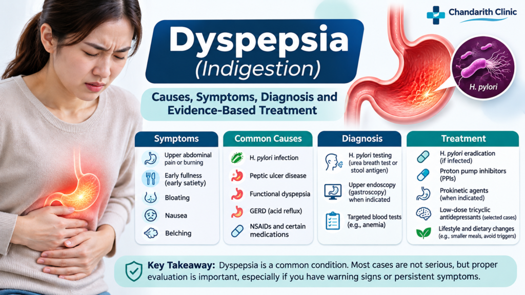
Dyspepsia, commonly known as indigestion, is a frequent gastrointestinal complaint characterized by discomfort or pain in the upper abdomen. It may occur after eating, during fasting, or throughout the day. Some patients experience early fullness, bloating, nausea, or excessive belching.
Dyspepsia can result from several conditions, including Helicobacter pylori (H. pylori) infection, peptic ulcer disease, gastroesophageal reflux disease (GERD), medication-related gastric irritation, and functional dyspepsia.
Although most cases are not caused by serious disease, persistent symptoms should be evaluated appropriately to exclude peptic ulcer disease and upper gastrointestinal malignancy.
This article explains the common causes, symptoms, diagnostic approach, treatment options, and prevention strategies based on contemporary clinical guidelines.
1. What is dyspepsia?
Dyspepsia refers to a group of symptoms originating from the upper gastrointestinal tract, particularly the stomach and duodenum.
The main symptoms include:
-
Epigastric pain or burning.
-
Postprandial fullness (uncomfortable fullness after meals).
-
Early satiety (feeling full after eating a small amount).
-
Upper abdominal bloating.
-
Nausea and belching.
Dyspepsia is generally classified into two major categories:
1. Organic dyspepsia
Symptoms are caused by an identifiable underlying disease, such as:
-
Peptic ulcer disease.
-
H. pylori-associated gastritis.
-
Gastric or duodenal malignancy.
-
Medication-induced gastropathy.
-
Other structural gastrointestinal diseases.
2. Functional dyspepsia
Symptoms occur without an identifiable structural explanation on appropriate investigation, including upper gastrointestinal endoscopy when indicated.
Functional dyspepsia is a disorder of gut-brain interaction and may involve impaired gastric accommodation, altered gastric emptying, visceral hypersensitivity, and changes in gut-brain signaling.
2. Common causes and risk factors
The most common causes of dyspepsia include:
| Cause | Clinical explanation |
|---|---|
| H. pylori infection | Chronic gastric infection associated with gastritis and peptic ulcer disease |
| Functional dyspepsia | Impaired gastric accommodation and visceral hypersensitivity |
| Peptic ulcer disease | Gastric or duodenal mucosal ulceration |
| GERD | Reflux of gastric contents into the esophagus |
| NSAIDs | Aspirin, ibuprofen, diclofenac, and naproxen may cause gastric injury |
| Smoking | Associated with gastrointestinal symptoms and ulcer complications |
| Alcohol | May aggravate upper gastrointestinal symptoms |
| Certain medications | Iron, potassium supplements, and some other drugs may cause dyspepsia |
Important: H. pylori infection and NSAID exposure are particularly important because both are associated with peptic ulcer disease.
3. Symptoms of dyspepsia
Symptoms can vary from mild intermittent discomfort to persistent upper abdominal pain.
Typical symptoms
-
Epigastric pain: discomfort or pain in the upper middle abdomen.
-
Epigastric burning: burning sensation in the stomach region.
-
Postprandial fullness: uncomfortable fullness after meals.
-
Early satiety: inability to finish a normal-sized meal.
-
Nausea: feeling sick or wanting to vomit.
-
Bloating: sensation of abdominal distension.
-
Belching: frequent release of gas from the stomach.
Clinical subtypes of functional dyspepsia
According to the Rome IV classification, functional dyspepsia has two major subtypes:
A. Postprandial Distress Syndrome (PDS)
-
Postprandial fullness after ordinary meals.
-
Early satiation that prevents finishing a regular meal.
-
Symptoms typically occur several times per week.
B. Epigastric Pain Syndrome (EPS)
-
Epigastric pain.
-
Epigastric burning.
-
Symptoms may occur during fasting or after meals.
Some patients have overlapping features of both subtypes.
4. When should you seek medical attention?
Most patients with dyspepsia do not have serious disease. However, certain symptoms require prompt medical evaluation.
Warning signs (alarm features)
-
Unintentional weight loss.
-
Persistent or recurrent vomiting.
-
Difficulty swallowing (dysphagia).
-
Painful swallowing (odynophagia).
-
Gastrointestinal bleeding, including vomiting blood or black stools.
-
Iron-deficiency anemia.
-
Palpable abdominal mass or lymphadenopathy.
-
Progressive symptoms, particularly in older patients.
-
Family history of upper gastrointestinal cancer.
Important: Alarm features should prompt clinical assessment, but they do not automatically mean cancer. The decision to perform endoscopy depends on age, severity, progression, family history, and individual cancer risk.
5. Diagnosis of dyspepsia
The diagnostic approach aims to identify the underlying cause, detect serious disease, and distinguish organic dyspepsia from functional dyspepsia.
A. Medical history
A detailed history should include:
-
Onset, duration, and frequency of symptoms.
-
Relationship between symptoms and meals.
-
Location and character of abdominal pain.
-
Presence of nausea, vomiting, reflux, or bloating.
-
Weight loss and appetite changes.
-
NSAID, aspirin, and other medication use.
-
Previous H. pylori infection or treatment.
-
Smoking and alcohol consumption.
-
Family history of gastric cancer.
B. Physical examination
Examination should include:
-
General appearance and nutritional status.
-
Vital signs.
-
Pallor and signs of anemia.
-
Abdominal examination for tenderness, distension, or masses.
-
Examination for lymphadenopathy or other concerning findings when indicated.
C. Laboratory investigations
Laboratory testing should be guided by clinical presentation.
| Investigation | Clinical indication |
|---|---|
| CBC | Suspected anemia or gastrointestinal bleeding |
| H. pylori stool antigen | Detection of active H. pylori infection |
| Urea breath test | Detection of active H. pylori infection |
| Liver function tests | Suspected hepatobiliary disease |
| Renal function | Relevant comorbidities or suspected systemic disease |
| Lipase | Suspected pancreatitis |
| Iron studies | Suspected iron-deficiency anemia |
Routine extensive laboratory testing is not necessary for every patient with uncomplicated dyspepsia.
D. H. pylori testing
Testing for H. pylori is an important part of dyspepsia management.
Preferred non-invasive tests include:
1. Urea breath test (UBT)
-
Highly accurate for active H. pylori infection.
-
Useful for initial diagnosis and confirmation of eradication.
2. Stool antigen test
-
Detects active infection.
-
Also suitable for confirmation of eradication.
3. Serology
-
Detects antibodies against H. pylori.
-
Cannot reliably distinguish current from previous infection.
-
Not recommended for confirming eradication.
Preparation before testing:
-
Stop proton pump inhibitors (PPIs) for at least 2 weeks before testing.
-
Avoid antibiotics and bismuth for at least 4 weeks before testing.
These precautions reduce false-negative results.
E. Upper gastrointestinal endoscopy (EGD)
EGD is useful for identifying:
-
Gastritis and erosive gastropathy.
-
Gastric and duodenal ulcers.
-
Esophagitis.
-
Gastric malignancy.
-
Other structural abnormalities.
Who should undergo endoscopy?
The ACG/CAG guideline recommends upper GI endoscopy for patients aged 60 years or older who present with new dyspepsia.
Endoscopy may be considered at a younger age when there are important risk factors, such as:
-
Family history of gastric cancer.
-
Childhood or prolonged residence in a high gastric cancer incidence region.
-
Significant alarm features.
-
Persistent or progressive symptoms despite appropriate treatment.
In Cambodia and other Asian countries where gastric cancer risk and H. pylori prevalence may differ from Western populations, local epidemiology should influence the threshold for investigation.
6. Treatment of dyspepsia
Treatment depends on the underlying cause.
The main objectives are to relieve symptoms, eradicate H. pylori when present, heal mucosal injury, and improve quality of life.
A. Lifestyle and dietary modifications
Lifestyle changes may improve symptoms, especially in functional dyspepsia.
Recommended measures include:
-
Eat smaller, more frequent meals.
-
Avoid overeating and very large meals.
-
Reduce fatty and fried foods if they trigger symptoms.
-
Limit coffee, tea, and carbonated drinks if symptoms worsen.
-
Avoid smoking.
-
Limit or avoid alcohol.
-
Avoid lying down immediately after meals.
-
Maintain a healthy body weight.
-
Manage stress and maintain adequate sleep.
-
Avoid unnecessary NSAID use.
There is no single diet that works for everyone. Patients should identify their individual food triggers.
B. H. pylori eradication therapy
All patients with confirmed H. pylori infection should generally receive eradication treatment unless there is a contraindication.
The 2024 ACG H. pylori guideline recommends optimized bismuth quadruple therapy for 14 days as the preferred empiric regimen when antibiotic susceptibility is unknown.
Optimized bismuth quadruple therapy – 14 days
| Medication | Dose |
|---|---|
| PPI (e.g., omeprazole) | 20 mg twice daily |
| Bismuth subcitrate | 120–300 mg four times daily, depending on formulation |
| Tetracycline | 500 mg four times daily |
| Metronidazole | 500 mg three or four times daily |
Use locally approved formulations and dosing. Bismuth subsalicylate has a different dose from bismuth subcitrate.
Important clinical considerations:
-
Avoid empiric clarithromycin triple therapy unless clarithromycin susceptibility is demonstrated.
-
Avoid empiric levofloxacin-based therapy unless susceptibility is confirmed.
-
Consider local antibiotic resistance, prior antibiotic exposure, allergies, and availability.
-
Ensure patients complete the entire treatment course.
Confirmation of eradication
A test of cure is recommended in all treated patients.
-
Perform urea breath test or stool antigen test at least 4 weeks after completing antibiotics.
-
Stop PPI therapy for at least 2 weeks before testing.
-
Avoid antibiotics and bismuth for at least 4 weeks before testing.
A negative test confirms successful eradication when appropriate testing conditions are followed.
C. Proton pump inhibitors (PPIs)
PPIs are first-line pharmacological treatment for patients who are H. pylori-negative or remain symptomatic after successful eradication.
Commonly used medications include:
| Medication | Typical adult dose |
|---|---|
| Omeprazole | 20 mg once daily |
| Esomeprazole | 20–40 mg once daily |
| Pantoprazole | 40 mg once daily |
| Lansoprazole | 30 mg once daily |
| Rabeprazole | 20 mg once daily |
Treatment approach:
-
Start with a standard-dose PPI once daily, usually before breakfast.
-
Assess response after 4–8 weeks.
-
If symptoms improve, consider stepping down or stopping treatment when appropriate.
-
Reassess the diagnosis if symptoms persist.
Long-term PPI treatment should be reserved for patients with a clear indication and periodically reviewed.
D. H2-receptor antagonists
H2-receptor antagonists may be considered when PPIs are unsuitable or when additional acid suppression is needed.
Examples include:
-
Famotidine.
-
Other locally available H2-receptor antagonists.
These medications reduce gastric acid secretion but may be less effective than PPIs for some patients.
E. Prokinetic agents
Prokinetic agents may be useful in patients with predominant postprandial fullness, early satiety, or suspected impaired gastric emptying.
Examples include:
-
Itopride.
-
Domperidone.
-
Metoclopramide.
-
Acotiamide, where available.
Clinical considerations:
-
Metoclopramide may cause extrapyramidal adverse effects and tardive dyskinesia; use the lowest effective dose for the shortest appropriate duration.
-
Domperidone may prolong the QT interval and increase the risk of serious arrhythmias.
-
Itopride may be considered where available, although evidence and regulatory approval vary.
Selection should depend on symptom pattern, availability, contraindications, and safety.
F. Tricyclic antidepressants (TCAs)
For patients with functional dyspepsia who do not respond adequately to PPI therapy and H. pylori eradication, low-dose tricyclic antidepressants may be considered.
Example:
-
Amitriptyline 10 mg at bedtime initially.
-
Increase gradually according to response and tolerability.
TCAs may improve visceral hypersensitivity and modulate gut-brain signaling.
Potential adverse effects include:
-
Dry mouth.
-
Constipation.
-
Drowsiness.
-
Urinary retention.
-
Cardiac conduction abnormalities.
Use caution in older adults and patients with cardiac disease or significant anticholinergic risk.
G. Psychological therapies
Functional dyspepsia is a disorder of gut-brain interaction. Psychological factors can contribute to symptom severity.
Selected patients may benefit from:
-
Cognitive behavioral therapy (CBT).
-
Stress management.
-
Relaxation techniques.
-
Gut-directed psychological interventions.
These approaches can be considered when symptoms persist despite standard medical treatment.
7. Management algorithm for dyspepsia
The following is a practical approach for clinicians based on ACG/CAG recommendations, H. pylori guidance, and updated Asian consensus recommendations.
Clinical approach to adult dyspepsia
Patient presents with dyspeptic symptoms
Epigastric pain/burning, early satiety or postprandial fullness
Step 1: Assess age, alarm features and cancer risk
-
Older age or high gastric cancer risk?
-
Bleeding, weight loss, progressive dysphagia, persistent vomiting or anemia?
Higher risk
Consider early EGD, with urgency determined by clinical findings.
Lower risk
Non-invasive H. pylori test-and-treat approach.
Step 2: H. pylori testing
-
Positive: Give appropriate eradication therapy and confirm cure.
-
Negative: Start standard-dose PPI for 4–8 weeks.
Step 3: Reassess clinical response
-
Improved: Step down or discontinue PPI when appropriate.
-
Persistent symptoms: Review adherence, diagnosis, medication triggers and indication for EGD.
Step 4: Consider functional dyspepsia treatment
-
Low-dose TCA.
-
Selected prokinetic therapy.
-
Psychological therapy and gut-brain interventions.
Clinical note: The age threshold of 60 years comes from the ACG/CAG guideline and is not an absolute rule for every Asian population. In regions with higher gastric cancer incidence, clinicians may reasonably consider earlier endoscopy. Alarm features require individualized assessment rather than automatic application of a single age cutoff.
8. Functional dyspepsia: Updated understanding
Functional dyspepsia is one of the most common causes of persistent dyspeptic symptoms.
It is classified as a disorder of gut-brain interaction rather than simply excessive gastric acid production.
Proposed mechanisms
-
Impaired gastric accommodation: the stomach does not relax appropriately after meals.
-
Delayed gastric emptying: food moves more slowly from the stomach in some patients.
-
Visceral hypersensitivity: increased sensitivity to normal gastric distension.
-
Duodenal abnormalities: low-grade inflammation and altered mucosal immune responses may contribute.
-
Gut-brain interaction: altered signaling between the gastrointestinal tract and central nervous system.
Updated management principles
The 2026 Second Asian Consensus Report emphasizes a more comprehensive, individualized approach to functional dyspepsia.
Key principles include:
-
Establish a positive clinical diagnosis rather than performing unnecessary investigations.
-
Identify and eradicate H. pylori when present.
-
Use acid suppression when appropriate.
-
Select treatment according to predominant symptoms.
-
Consider neuromodulators for persistent symptoms.
-
Address dietary factors, psychological stress, and quality of life.
-
Avoid unnecessary investigations and medications without a clear indication.
Evidence for emerging therapies, including probiotics and potassium-competitive acid blockers (P-CABs), remains variable and should be interpreted according to the specific clinical indication.
PMC+1
9. Differential diagnosis
Not every patient with epigastric discomfort has dyspepsia caused by gastric disease.
Important differential diagnoses include:
| Condition | Suggestive clinical features |
|---|---|
| GERD | Heartburn and acid regurgitation |
| Peptic ulcer disease | Epigastric pain, possible bleeding |
| Gastric cancer | Progressive symptoms, weight loss, anemia |
| Gallstone disease | Right upper quadrant or postprandial pain |
| Acute pancreatitis | Severe persistent epigastric pain, often radiating to the back |
| Chronic pancreatitis | Recurrent pain, weight loss, steatorrhea |
| Biliary disease | Right upper abdominal pain, jaundice when obstructive |
| Myocardial ischemia | Epigastric discomfort with exertion or cardiovascular risk factors |
| Gastroparesis | Early satiety, nausea, vomiting and delayed gastric emptying |
In patients with diabetes, consider diabetic gastroparesis when early satiety, nausea, and vomiting are prominent.
10. Possible complications
Uncomplicated functional dyspepsia does not usually cause structural gastrointestinal complications.
However, underlying organic diseases may lead to serious complications.
Examples include:
-
Peptic ulcer bleeding.
-
Gastric or duodenal perforation.
-
Gastric outlet obstruction.
-
Iron-deficiency anemia from chronic gastrointestinal blood loss.
-
Gastric malignancy in patients with underlying cancer.
Persistent or worsening symptoms should not simply be treated with repeated courses of acid-suppressive medication without reassessment.
11. Prevention of dyspepsia
Patients can reduce the frequency and severity of symptoms through several practical measures:
-
Maintain regular eating habits.
-
Eat moderate-sized meals.
-
Avoid foods that consistently trigger symptoms.
-
Reduce excessive coffee consumption if symptomatic.
-
Avoid smoking and excessive alcohol.
-
Avoid unnecessary NSAIDs.
-
Maintain a healthy body weight.
-
Manage stress and sleep adequately.
-
Seek evaluation for persistent upper abdominal symptoms.
H. pylori detection and eradication are particularly important in patients with confirmed infection.
12. Frequently asked questions (FAQ)
1. What is the most common cause of dyspepsia?
Common causes include functional dyspepsia, H. pylori infection, peptic ulcer disease, GERD, and medication-related gastric irritation. The underlying cause varies between populations.
2. Is dyspepsia the same as gastritis?
No. Dyspepsia is a group of symptoms, whereas gastritis is inflammation of the gastric mucosa. A patient can have dyspepsia without gastritis, and gastritis may occur without symptoms.
3. Can H. pylori cause dyspepsia?
Yes. H. pylori can cause chronic gastritis and peptic ulcer disease and may contribute to functional dyspepsia symptoms. Eradication can improve symptoms in some infected patients.
4. Can stress cause dyspepsia?
Stress and psychological factors can influence gut-brain signaling, gastric sensitivity, and symptom perception. They may aggravate functional dyspepsia.
5. What is the best medicine for dyspepsia?
There is no single best medicine for every patient.
-
H. pylori-positive: appropriate eradication therapy.
-
H. pylori-negative: consider a PPI trial.
-
Persistent functional dyspepsia: consider a low-dose TCA or selected prokinetic agent.
6. How long should patients take PPIs?
For uncomplicated dyspepsia, a typical initial trial is 4–8 weeks. Continued therapy should depend on the clinical response and underlying diagnosis.
7. Can dyspepsia cause bloating?
Yes. Bloating, belching, and postprandial fullness are common associated symptoms, especially in functional dyspepsia.
8. Is dyspepsia dangerous?
Most cases are not dangerous. However, persistent symptoms, gastrointestinal bleeding, unexplained weight loss, progressive swallowing difficulty, or persistent vomiting require medical assessment.
9. When is gastroscopy necessary?
Endoscopy is generally recommended for new dyspepsia in patients aged 60 years or older under ACG/CAG guidance. Younger patients may also need endoscopy depending on cancer risk, alarm features, and treatment response.
10. Can dyspepsia be cured permanently?
Many patients achieve substantial or complete symptom relief. H. pylori eradication may resolve symptoms in some patients, while functional dyspepsia can have a fluctuating course requiring individualized long-term management.
11. Can patients with dyspepsia drink coffee or milk?
Some patients tolerate these beverages, while others experience worsening symptoms. Patients should identify individual triggers rather than follow unnecessarily restrictive diets.
12. Does dyspepsia increase the risk of stomach cancer?
Dyspepsia itself does not cause stomach cancer. However, H. pylori infection and certain gastric premalignant conditions are associated with increased gastric cancer risk. Persistent or concerning symptoms should be investigated appropriately.
13. Key clinical take-home messages
For healthcare professionals, the following points are particularly important:
-
Dyspepsia is a symptom complex, not a definitive diagnosis.
-
Always consider H. pylori infection and NSAID-associated mucosal injury.
-
Use a risk-based approach to decide who needs upper GI endoscopy.
-
Test-and-treat for H. pylori is an important first-line strategy in appropriate younger patients.
-
Use PPIs for patients who are H. pylori-negative or remain symptomatic after eradication.
-
Consider functional dyspepsia when no explanatory structural disease is identified.
-
For persistent functional dyspepsia, consider TCAs, selected prokinetics, and gut-brain interventions.
-
Avoid repeated empirical treatment without reassessing the diagnosis.
14. References and clinical guidelines
The following references support the diagnostic and treatment recommendations in this article.
1. Moayyedi PM, Lacy BE, Andrews CN, Enns RA, Howden CW, Vakil N. ACG and CAG Clinical Guideline: Management of Dyspepsia. American Journal of Gastroenterology. 2017;112(7):988–1013.
DOI: 10.1038/ajg.2017.154
2. Chey WD, Howden CW, Moss SF, et al. ACG Clinical Guideline: Treatment of Helicobacter pylori Infection. American Journal of Gastroenterology. 2024;119(9):1730–1753.
DOI: 10.14309/ajg.0000000000002968
3. Black CJ, Paine PA, Agrawal A, et al. British Society of Gastroenterology guidelines on the management of functional dyspepsia. Gut. 2022;71:1697–1723.
DOI: 10.1136/gutjnl-2022-327737
4. Mahadeva S, et al. Second Asian Consensus Report on Functional Dyspepsia (2025): Updated Recommendations. Journal of Gastroenterology and Hepatology. 2026;41(3):832–853.
DOI: 10.1111/jgh.70222
5. Drossman DA, Hasler WL. Rome IV—Functional GI Disorders: Disorders of Gut-Brain Interaction. Gastroenterology. 2016;150(6):1257–1261.
DOI: 10.1053/j.gastro.2016.03.035
6. American College of Gastroenterology. ACG and CAG Clinical Guideline: Management of Dyspepsia.
Official ACG guideline resource
Medical disclaimer: This article is intended for educational purposes and does not replace an individual medical consultation. Diagnosis and treatment should be determined by a qualified healthcare professional.