
Medical Review: Dr. Cheang Chandarith, Internist
Introduction
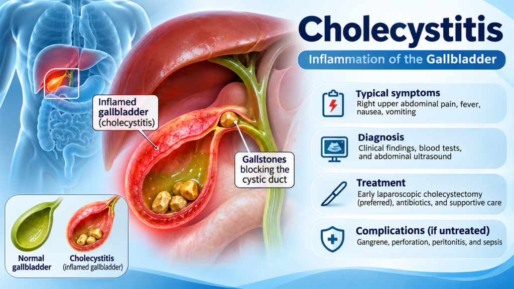
Cholecystitis is inflammation of the gallbladder, a small pear-shaped organ located beneath the liver in the right upper abdomen. The gallbladder stores and concentrates bile, which helps the body digest dietary fats.
The most common cause of acute cholecystitis is obstruction of the cystic duct by gallstones. This obstruction causes bile retention, increased pressure inside the gallbladder, inflammation, and sometimes secondary bacterial infection.
Acute cholecystitis is an important cause of acute abdominal pain and requires prompt medical assessment. Without appropriate treatment, it may progress to serious complications, including gallbladder gangrene, perforation, peritonitis, and sepsis.
Early diagnosis, appropriate antibiotic therapy when indicated, and timely surgical management are essential to improving patient outcomes.
1. Types of Cholecystitis
Cholecystitis can be classified into several types.
1.1 Acute calculous cholecystitis
Acute calculous cholecystitis accounts for approximately 90–95% of acute cases.
It develops when a gallstone obstructs the cystic duct, resulting in:
- Bile retention and gallbladder distension.
- Increased intraluminal pressure.
- Impaired gallbladder wall circulation.
- Local inflammatory response.
- Possible secondary bacterial infection.
Common associated bacteria include:
- Escherichia coli
- Klebsiella species
- Enterococcus species
- Enterobacter species
- Other Enterobacterales and anaerobic bacteria in selected complicated infections.
1.2 Acute acalculous cholecystitis
Acalculous cholecystitis occurs without gallstones and accounts for approximately 5–10% of acute cholecystitis cases.
It is commonly associated with critically ill patients and may develop due to gallbladder ischemia, bile stasis, and systemic inflammation.
Risk factors include:
- Severe sepsis or septic shock.
- Major surgery.
- Severe trauma or burns.
- Prolonged fasting or total parenteral nutrition.
- Mechanical ventilation.
- Severe cardiovascular disease.
- Immunosuppression.
Acalculous cholecystitis can progress rapidly to gangrene and perforation. Clinical suspicion is particularly important in critically ill patients who cannot communicate abdominal symptoms.
1.3 Chronic cholecystitis
Chronic cholecystitis usually results from repeated episodes of gallbladder inflammation, most commonly associated with gallstones.
Long-standing inflammation can cause:
- Gallbladder wall fibrosis.
- Thickening of the gallbladder wall.
- Reduced gallbladder contractility.
- Recurrent right upper quadrant discomfort.
- Chronic digestive symptoms.
2. Causes and Risk Factors
Gallstones are the principal cause of acute calculous cholecystitis.
Important risk factors for gallstone formation include:
- Female sex.
- Increasing age.
- Obesity.
- Rapid weight loss.
- Pregnancy.
- Family history of gallstones.
- Diabetes mellitus.
- Metabolic syndrome.
- Certain medications, including some estrogen-containing therapies.
- Liver cirrhosis and hemolytic disorders.
However, gallstones do not always cause symptoms. Many patients have asymptomatic cholelithiasis and never develop cholecystitis.
3. Clinical Symptoms of Cholecystitis
The typical presentation of acute cholecystitis includes:
3.1 Right upper quadrant abdominal pain
Pain is usually located in the right upper abdomen and may radiate to the right shoulder or back.
Characteristics include:
- Persistent pain lasting more than 4–6 hours.
- Progressive pain intensity.
- Pain aggravated by deep inspiration or movement.
- Localized tenderness over the gallbladder.
Unlike uncomplicated biliary colic, acute cholecystitis usually produces persistent inflammatory pain.
3.2 Fever
Fever is a common manifestation of systemic inflammation and may indicate associated infection.
3.3 Nausea and vomiting
Patients frequently experience nausea, vomiting, loss of appetite, and abdominal discomfort.
3.4 Murphy’s sign
Murphy’s sign is an important physical examination finding.
It is positive when the patient experiences sudden pain and inspiratory arrest during deep inspiration while the examiner palpates the right upper quadrant.
3.5 Other clinical findings
Additional symptoms and signs may include:
- Abdominal guarding.
- Right upper quadrant tenderness.
- Leukocytosis.
- Elevated C-reactive protein (CRP).
- Mild elevation of liver enzymes.
Jaundice is not typical of isolated acute cholecystitis. Its presence should raise suspicion of common bile duct obstruction, choledocholithiasis, or associated cholangitis.
4. Diagnosis of Acute Cholecystitis
Diagnosis is based on clinical findings, laboratory investigations, and imaging studies.
The Tokyo Guidelines 2018 (TG18) provide widely used diagnostic criteria.
4.1 Tokyo Guidelines diagnostic criteria
Three categories of findings are considered:
A. Local signs of inflammation
- Murphy’s sign.
- Right upper quadrant pain, tenderness, or mass.
B. Systemic signs of inflammation
- Fever.
- Elevated white blood cell count.
- Elevated CRP.
C. Imaging findings
Characteristic imaging findings of acute cholecystitis.
Diagnostic classification:
- Suspected diagnosis: At least one local sign (A) and one systemic sign (B).
- Definite diagnosis: At least one local sign (A), one systemic sign (B), and characteristic imaging findings (C).
Other causes of acute abdominal pain should be excluded.
4.2 Laboratory investigations
Recommended investigations include:
| Investigation | Clinical significance |
|---|---|
| CBC | Leukocytosis and neutrophilia |
| CRP | Systemic inflammation |
| AST and ALT | Assess hepatocellular injury |
| ALP and GGT | Evaluate cholestasis |
| Total and direct bilirubin | Assess possible biliary obstruction |
| Renal function | Assess dehydration and organ dysfunction |
| Electrolytes | Assess metabolic abnormalities |
| Lipase | Exclude acute pancreatitis |
| Blood cultures | Consider in sepsis or severe infection |
Liver function tests may be normal or mildly elevated in uncomplicated acute cholecystitis.
Marked bilirubin elevation or significant cholestatic abnormalities should prompt evaluation for common bile duct stones or cholangitis.
4.3 Imaging studies
A. Abdominal ultrasound
Ultrasound is the first-line imaging investigation.
Typical findings include:
- Gallstones.
- Gallbladder wall thickening (usually greater than 3–4 mm).
- Gallbladder distension.
- Pericholecystic fluid.
- Sonographic Murphy’s sign.
- Increased vascularity of the gallbladder wall.
Ultrasound is inexpensive, widely available, and does not expose patients to ionizing radiation.
B. Computed tomography (CT)
CT is useful when:
- Ultrasound findings are inconclusive.
- Complications are suspected.
- Alternative abdominal diagnoses need to be excluded.
- Gangrenous or emphysematous cholecystitis is suspected.
CT may demonstrate gallbladder wall thickening, surrounding inflammatory changes, abscess formation, perforation, or gas within the gallbladder wall.
C. Magnetic resonance imaging (MRI/MRCP)
MRI and MRCP are useful when:
- Ultrasound is inconclusive.
- Common bile duct stones are suspected.
- There is unexplained jaundice or abnormal liver function.
- Further biliary tract evaluation is necessary.
D. Hepatobiliary scintigraphy (HIDA scan)
HIDA scanning is useful when acute cholecystitis remains suspected despite inconclusive ultrasound findings.
Failure of the gallbladder to visualize during the examination supports cystic duct obstruction.
5. Severity Grading of Acute Cholecystitis
The Tokyo Guidelines 2018 classify acute cholecystitis into three grades.
Grade I: Mild acute cholecystitis
- Does not meet Grade II or Grade III criteria.
- No organ dysfunction.
- Usually responds well to early surgical treatment.
Grade II: Moderate acute cholecystitis
Any of the following:
- WBC greater than 18,000/mm³.
- Palpable tender mass in the right upper quadrant.
- Symptoms lasting more than 72 hours.
- Marked local inflammation, such as gangrenous cholecystitis, pericholecystic abscess, hepatic abscess, biliary peritonitis, or emphysematous cholecystitis.
Grade III: Severe acute cholecystitis
Associated with dysfunction of at least one organ system:
- Cardiovascular: Hypotension requiring dopamine ≥5 μg/kg/min or any dose of norepinephrine.
- Neurological: Decreased level of consciousness.
- Respiratory: PaO₂/FiO₂ ratio below 300.
- Renal: Oliguria or creatinine greater than 2.0 mg/dL.
- Hepatic: PT-INR greater than 1.5.
- Hematological: Platelet count below 100,000/mm³.
Severity grading is important for determining the appropriate treatment strategy and need for intensive care.
6. Treatment of Acute Cholecystitis
Management depends on disease severity, the patient’s general condition, associated complications, and surgical risk.
6.1 Initial medical management
Initial treatment includes:
1. Hospital admission
Patients with suspected acute cholecystitis generally require hospital assessment and surgical consultation.
2. Fasting
Keep patients nil by mouth initially, particularly when surgery is anticipated or vomiting is present.
3. Intravenous fluid therapy
Administer isotonic crystalloid fluids according to the patient’s hydration status, hemodynamic condition, renal function, and cardiac status.
4. Pain control
Common options include:
- NSAIDs when appropriate.
- Paracetamol.
- Opioid analgesics for severe pain when necessary.
5. Antiemetics
Use antiemetic medications when nausea and vomiting are significant.
6. Monitoring
Monitor:
- Blood pressure and heart rate.
- Temperature.
- Urine output.
- Renal function.
- Electrolytes.
- Signs of sepsis or organ dysfunction.
6.2 Antibiotic therapy
Antibiotics are indicated for acute cholecystitis, particularly when systemic inflammation, bacterial infection, or complicated disease is present.
Empirical antibiotic selection should consider disease severity, community versus healthcare-associated infection, local antimicrobial resistance patterns, previous antibiotic exposure, renal function, and risk of resistant organisms.
Common empirical options include:
Mild to moderate community-acquired infection
- Ceftriaxone 1–2 g IV every 24 hours, with metronidazole 500 mg IV every 8–12 hours when anaerobic coverage is indicated.
- Amoxicillin-clavulanate IV according to local susceptibility and institutional protocols.
- Other appropriate beta-lactam regimens according to local resistance patterns.
Severe infection or healthcare-associated infection
- Piperacillin-tazobactam 4.5 g IV every 6–8 hours, adjusted according to renal function and infusion protocol.
- Cefepime plus metronidazole when appropriate.
- Carbapenems, such as meropenem, when there is a significant risk of ESBL-producing organisms or other resistant pathogens.
Additional coverage for Enterococcus or MRSA should be considered only when specific clinical risk factors or microbiological evidence justify it.
Duration of antibiotic therapy
Antibiotic duration should depend on disease severity, clinical response, and adequacy of source control.
- After uncomplicated acute cholecystitis with successful cholecystectomy and adequate source control, routine postoperative antibiotics are generally unnecessary.
- Complicated infection, gangrene, perforation, abscess, or persistent sepsis may require a longer course.
- For complicated intra-abdominal infection with adequate source control, a short course of approximately 4 days is often sufficient, with individualization according to clinical progress.
Antibiotics do not replace definitive surgical management.
6.3 Surgical treatment: Laparoscopic cholecystectomy
Early laparoscopic cholecystectomy is the preferred definitive treatment for most patients with acute calculous cholecystitis who can tolerate surgery.
Benefits include:
- Removal of the source of inflammation.
- Prevention of recurrent cholecystitis.
- Reduced risk of recurrent biliary complications.
- Shorter hospital stay.
- Earlier recovery.
Timing of surgery
Early laparoscopic cholecystectomy should be performed as soon as reasonably possible, preferably within 72 hours of symptom onset when feasible.
Current practice supports early surgery during the index admission rather than routinely delaying surgery for several weeks.
The exact timing depends on patient stability, disease severity, surgical expertise, and available facilities.
6.4 Percutaneous cholecystostomy
Percutaneous cholecystostomy involves placing a drainage catheter into the gallbladder under ultrasound or CT guidance.
It may be considered in selected patients with:
- Severe acute cholecystitis.
- Uncontrolled infection.
- Significant organ dysfunction.
- High operative risk.
- Inability to undergo immediate surgery.
It provides temporary gallbladder drainage and source control.
However, it should not automatically replace cholecystectomy in patients who are high-risk but still suitable for surgery. The CHOCOLATE trial demonstrated better outcomes with laparoscopic cholecystectomy than percutaneous drainage in selected high-risk patients.
The decision should be individualized with surgical and interventional radiology input.
7. Complications of Acute Cholecystitis
Delayed diagnosis or inadequate treatment may lead to serious complications.
7.1 Gallbladder empyema
Accumulation of pus within the gallbladder due to bacterial infection.
7.2 Gangrenous cholecystitis
Severe inflammation and ischemia cause necrosis of the gallbladder wall.
It is associated with an increased risk of perforation and sepsis.
7.3 Gallbladder perforation
Perforation may result in localized abscess formation or generalized biliary peritonitis.
7.4 Emphysematous cholecystitis
A severe form characterized by gas-forming bacterial infection within the gallbladder wall or lumen.
It is more common in patients with diabetes mellitus and has a high risk of gangrene and perforation.
7.5 Sepsis and septic shock
Severe infection may lead to systemic organ dysfunction, hypotension, and life-threatening septic shock.
7.6 Biliary complications
Associated complications include:
- Choledocholithiasis.
- Acute cholangitis.
- Gallstone pancreatitis.
- Mirizzi syndrome.
8. Differential Diagnosis
Right upper quadrant abdominal pain is not specific to cholecystitis.
Important differential diagnoses include:
- Biliary colic.
- Choledocholithiasis.
- Acute cholangitis.
- Acute pancreatitis.
- Peptic ulcer disease.
- Acute hepatitis.
- Liver abscess.
- Right-sided pyelonephritis.
- Right lower lobe pneumonia.
- Myocardial ischemia, particularly in older patients and patients with diabetes.
Clinical examination, laboratory tests, and appropriate imaging are important for distinguishing these conditions.
9. Prevention of Gallstones and Cholecystitis
Although not all gallstones can be prevented, several lifestyle measures may reduce the risk.
Recommendations include:
- Maintain a healthy body weight.
- Avoid rapid weight loss.
- Exercise regularly.
- Follow a balanced diet rich in vegetables and fiber.
- Reduce excessive intake of refined carbohydrates.
- Limit saturated fats.
- Control diabetes and metabolic risk factors.
Patients with symptomatic gallstones should receive medical evaluation to determine whether elective cholecystectomy is indicated.
Routine prophylactic cholecystectomy is not recommended for most patients with asymptomatic gallstones.
10. When Should Patients Seek Emergency Medical Attention?
Patients should seek urgent medical attention if they experience:
- Persistent right upper abdominal pain.
- Fever with abdominal pain.
- Repeated vomiting.
- Yellow discoloration of the eyes or skin.
- Dark urine or pale stools.
- Severe abdominal tenderness.
- Confusion, fainting, or marked weakness.
- Low blood pressure or difficulty breathing.
These symptoms may indicate acute cholecystitis, biliary obstruction, cholangitis, or another serious abdominal condition.
11. Frequently Asked Questions (FAQ)
1. What is the main cause of cholecystitis?
Gallstones obstructing the cystic duct are the most common cause. This obstruction leads to bile retention, increased gallbladder pressure, and inflammation.
2. What is the difference between cholecystitis and gallstones?
Gallstones are solid deposits that form inside the gallbladder. Cholecystitis is inflammation of the gallbladder, most commonly caused by gallstones obstructing the cystic duct.
Not everyone with gallstones develops cholecystitis.
3. What are the typical symptoms of acute cholecystitis?
The most common symptoms are persistent right upper abdominal pain, fever, nausea, vomiting, and tenderness over the gallbladder. Pain may radiate to the right shoulder or back.
4. How is cholecystitis diagnosed?
Diagnosis is based on clinical symptoms, physical examination, inflammatory markers, and imaging. Abdominal ultrasound is generally the first-line imaging investigation.
5. Can cholecystitis resolve without surgery?
Some patients may initially improve with antibiotics, intravenous fluids, and supportive treatment. However, in suitable patients with acute calculous cholecystitis, laparoscopic cholecystectomy is the preferred definitive treatment to prevent recurrence and further complications.
6. Are antibiotics always necessary for cholecystitis?
Antibiotics are generally used in acute cholecystitis, especially when systemic inflammation or infection is present. The choice and duration depend on severity, local bacterial resistance, and adequacy of source control. Routine postoperative antibiotics are usually unnecessary after uncomplicated cholecystectomy with adequate source control.
7. What is the best treatment for acute cholecystitis?
Early laparoscopic cholecystectomy is the preferred definitive treatment for most patients who can tolerate surgery. Antibiotics and supportive care are important components of initial management.
8. How long does recovery take after laparoscopic cholecystectomy?
Many patients recover sufficiently to resume light daily activities within approximately 1–2 weeks. Recovery may take longer following complicated disease or open surgery.
9. Can a person live normally without a gallbladder?
Yes. Most people can live healthy lives without a gallbladder. Bile flows directly from the liver into the small intestine after gallbladder removal.
Some patients experience temporary changes in bowel habits, particularly diarrhea after fatty meals.
10. Is cholecystitis dangerous?
Yes. Although many cases respond well to appropriate treatment, delayed management may cause gallbladder gangrene, perforation, peritonitis, and sepsis.
11. What is the difference between acute calculous and acalculous cholecystitis?
Calculous cholecystitis is caused by gallstone obstruction of the cystic duct. Acalculous cholecystitis occurs without gallstones and is commonly associated with critically ill or hospitalized patients.
12. When is percutaneous cholecystostomy indicated?
It may be considered for patients with severe acute cholecystitis who cannot undergo immediate surgery or require temporary gallbladder drainage. It is not automatically the preferred treatment for every high-risk surgical patient.
Conclusion
Cholecystitis is a potentially serious inflammatory condition of the gallbladder, most commonly caused by gallstones obstructing the cystic duct.
Early recognition, appropriate diagnostic imaging, severity assessment, supportive treatment, and timely laparoscopic cholecystectomy are essential for optimal patient outcomes.
Clinicians should recognize high-risk patients early, identify complications such as gangrene and perforation, and ensure appropriate source control.
Early surgical consultation and individualized management remain the cornerstones of effective treatment.
References
- Yokoe M, Hata J, Takada T, et al. Tokyo Guidelines 2018: Diagnostic criteria and severity grading of acute cholecystitis. Journal of Hepato-Biliary-Pancreatic Sciences. 2018;25(1):41–54. doi:10.1002/jhbp.515.
- Okamoto K, Suzuki K, Takada T, et al. Tokyo Guidelines 2018: Flowchart for the management of acute cholecystitis. Journal of Hepato-Biliary-Pancreatic Sciences. 2018;25(1):55–72. doi:10.1002/jhbp.516.
- Pisano M, Allievi N, Gurusamy K, et al. 2020 World Society of Emergency Surgery updated guidelines for the diagnosis and treatment of acute calculus cholecystitis. World Journal of Emergency Surgery. 2020;15:61. doi:10.1186/s13017-020-00336-x.
- de’Angelis N, et al. Safe Cholecystectomy Multi-Society Practice Guideline and State of the Art Consensus Conference on Prevention of Bile Duct Injury during Cholecystectomy. Society of American Gastrointestinal and Endoscopic Surgeons (SAGES), 2020.
- Loozen CS, et al. Laparoscopic cholecystectomy versus percutaneous catheter drainage for acute cholecystitis in high-risk patients (CHOCOLATE). BMJ. 2018;363. doi:10.1136/bmj.k3965.
- Eastern Association for the Surgery of Trauma (EAST). Acute cholecystitis in high-risk patients: A systematic review, meta-analysis, and practice management guideline. Trauma Surgery & Acute Care Open. 2026. doi:10.1136/tsaco-2025-002191.
- SAGES. Guidelines for the Clinical Application of Laparoscopic Biliary Tract Surgery. Society of American Gastrointestinal and Endoscopic Surgeons.
Medical disclaimer: This article is intended for educational purposes and does not replace individual medical assessment, diagnosis, or treatment. Antibiotic selection and surgical decisions should follow local clinical guidelines, antimicrobial susceptibility patterns, and specialist recommendations.