
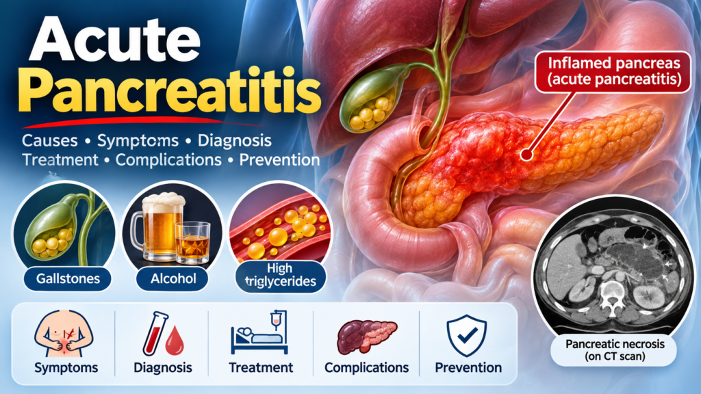
Acute pancreatitis is a sudden inflammatory condition of the pancreas that can range from mild, self-limiting disease to severe, life-threatening illness associated with organ failure, pancreatic necrosis, and systemic complications.
Early diagnosis, appropriate fluid resuscitation, effective pain control, early nutritional support, and identification of the underlying cause are essential to improving patient outcomes.
This article reviews the clinical presentation, diagnostic criteria, severity classification, investigations, evidence-based management, complications, and prevention of acute pancreatitis, drawing on major international guidelines, including the American College of Gastroenterology (ACG) 2024 guideline.
1. What is acute pancreatitis?
Acute pancreatitis is an acute inflammatory disorder caused by premature activation of digestive enzymes within the pancreas, resulting in pancreatic tissue injury and inflammation.
Normally, the pancreas produces digestive enzymes that become activated in the small intestine. In acute pancreatitis, abnormal intracellular activation of enzymes, particularly trypsin, contributes to pancreatic injury, inflammation, and sometimes necrosis.
The inflammatory response may remain localized to the pancreas or extend throughout the body, causing systemic inflammatory response syndrome (SIRS), respiratory failure, acute kidney injury, and circulatory shock.
Clinical spectrum:
-
Mild acute pancreatitis: No organ failure or local/systemic complications.
-
Moderately severe acute pancreatitis: Transient organ failure lasting less than 48 hours and/or local or systemic complications without persistent organ failure.
-
Severe acute pancreatitis: Persistent organ failure lasting more than 48 hours.
2. Causes of acute pancreatitis
The two most common causes are gallstones and alcohol consumption.
Major causes
| Cause | Clinical significance |
|---|---|
| Gallstones | One of the leading causes; stones may obstruct the ampulla of Vater |
| Alcohol | Usually associated with prolonged or substantial alcohol exposure |
| Hypertriglyceridemia | Particularly when triglycerides exceed 1,000 mg/dL |
| Medications | Certain drugs can cause drug-induced pancreatitis |
| Post-ERCP | Can occur following endoscopic retrograde cholangiopancreatography |
| Hypercalcemia | Increased calcium may contribute to pancreatic enzyme activation |
| Trauma | Abdominal injury or pancreatic trauma |
| Autoimmune pancreatitis | An uncommon cause |
| Genetic factors | Particularly in recurrent or unexplained cases |
Other possible causes include pancreatic tumors, anatomical abnormalities, infections, and idiopathic disease.
Important clinical consideration: In patients older than 40 years with unexplained acute pancreatitis, consider investigating for pancreatic malignancy, particularly when episodes recur or there is unexplained weight loss.
3. Symptoms and clinical presentation
The typical presentation includes:
-
Sudden onset of severe epigastric abdominal pain.
-
Pain radiating to the back.
-
Persistent pain lasting several hours or days.
-
Nausea and repeated vomiting.
-
Abdominal tenderness and distension.
-
Reduced bowel sounds.
-
Fever and tachycardia.
-
Dehydration and hypotension in severe cases.
Clinical signs of severe disease
-
Hypotension or shock.
-
Tachypnea and hypoxemia.
-
Oliguria or acute kidney injury.
-
Altered mental status.
-
Persistent SIRS.
-
Abdominal distension and ileus.
Rarely, severe hemorrhagic pancreatitis may produce:
-
Cullen sign: Periumbilical ecchymosis.
-
Grey Turner sign: Flank ecchymosis.
These signs are uncommon and generally indicate advanced disease.
4. Diagnosis of acute pancreatitis
The diagnosis requires at least two of the following three criteria:
-
Typical abdominal pain consistent with acute pancreatitis.
-
Serum lipase or amylase at least three times the upper limit of normal.
-
Characteristic findings of acute pancreatitis on abdominal imaging.
Laboratory investigations
| Investigation | Clinical purpose |
|---|---|
| Serum lipase | Preferred pancreatic enzyme test |
| Serum amylase | Alternative diagnostic enzyme |
| CBC | Leukocytosis, hematocrit |
| BUN and creatinine | Assess hydration and renal function |
| Electrolytes | Detect electrolyte abnormalities |
| Blood glucose | Assess hyperglycemia |
| Liver function tests | Identify possible biliary etiology |
| Serum triglycerides | Investigate hypertriglyceridemia |
| Serum calcium | Identify hypercalcemia |
| CRP | Assess inflammatory response and prognosis |
Lipase is preferred over amylase because it generally has better diagnostic performance and remains elevated longer.
Imaging investigations
1. Abdominal ultrasound
Recommended early in most patients to assess:
-
Gallstones.
-
Gallbladder inflammation.
-
Common bile duct dilation.
-
Possible biliary obstruction.
2. Contrast-enhanced CT scan
CT is not routinely required at admission when the diagnosis is already established clinically and biochemically.
Consider CT when:
-
The diagnosis is uncertain.
-
The patient fails to improve after 48–72 hours.
-
Complications such as necrosis, collections, or hemorrhage are suspected.
3. MRI/MRCP
Useful when evaluating suspected common bile duct stones, pancreatic duct abnormalities, or biliary obstruction, especially when ultrasound is inconclusive.
5. Severity classification: Revised Atlanta Classification
The Revised Atlanta Classification divides acute pancreatitis into three categories according to organ failure and complications.
| Classification | Definition |
|---|---|
| Mild | No organ failure and no local or systemic complications |
| Moderately severe | Transient organ failure (less than 48 hours) and/or local or systemic complications |
| Severe | Persistent organ failure lasting more than 48 hours |
Organ failure assessment
The modified Marshall scoring system evaluates three organ systems:
-
Respiratory system: PaO₂/FiO₂ ratio.
-
Renal system: Serum creatinine.
-
Cardiovascular system: Systolic blood pressure and response to fluid resuscitation.
A score of 2 or more in any one organ system indicates organ failure.
Predicting severity
Several clinical tools help identify patients at increased risk of severe disease.
BISAP score (Bedside Index for Severity in Acute Pancreatitis)
One point is assigned for each of the following:
| Parameter | Criteria |
|---|---|
| B | BUN greater than 25 mg/dL |
| I | Impaired mental status |
| S | SIRS present |
| A | Age greater than 60 years |
| P | Pleural effusion |
A BISAP score of 3 or higher indicates increased risk of severe disease and mortality. It should support, not replace, clinical assessment and repeated evaluation.
Systemic Inflammatory Response Syndrome (SIRS)
SIRS is present when at least two of the following criteria are met:
-
Temperature greater than 38°C or less than 36°C.
-
Heart rate greater than 90 beats/minute.
-
Respiratory rate greater than 20/minute or PaCO₂ less than 32 mmHg.
-
WBC greater than 12,000/mm³, less than 4,000/mm³, or greater than 10% immature bands.
Persistent SIRS, particularly at 48 hours, is associated with a higher risk of severe pancreatitis.
Important: No single scoring system can reliably predict the entire clinical course. Continuous reassessment during the first 48 hours is essential.
6. Treatment and management of acute pancreatitis
The primary goals of treatment are:
-
Restore and maintain adequate circulating volume.
-
Control abdominal pain.
-
Maintain adequate nutrition.
-
Identify and treat the underlying cause.
-
Prevent and manage organ failure and complications.
6.1 Initial assessment and stabilization
The initial management follows an ABCDE approach.
Airway and breathing
-
Assess oxygen saturation and respiratory status.
-
Administer oxygen if hypoxemic.
-
Monitor for acute respiratory distress syndrome (ARDS).
Circulation
-
Assess blood pressure, heart rate, capillary refill, and peripheral perfusion.
-
Establish intravenous access.
-
Begin appropriate intravenous crystalloid resuscitation.
-
Monitor urine output and renal function.
Monitoring
-
Vital signs and oxygen saturation.
-
Fluid input and output.
-
Urine output.
-
BUN, creatinine, hematocrit, electrolytes, and glucose.
-
Signs of persistent SIRS and organ failure.
Patients with shock, persistent organ failure, or significant respiratory or renal dysfunction require higher-level monitoring and potentially ICU admission.
6.2 Intravenous fluid resuscitation
Fluid resuscitation is one of the most important aspects of early management.
Preferred fluid: Lactated Ringer’s solution (Hartmann’s solution).
Current international recommendations favor moderate, goal-directed fluid resuscitation rather than routine aggressive fluid administration.
Suggested starting approach:
| Clinical situation | Fluid strategy |
|---|---|
| Most patients | Lactated Ringer’s approximately 1.5 mL/kg/hour |
| Hypovolemia | Consider 10 mL/kg IV bolus over approximately 2 hours |
| Hypotension or shock | Individualized resuscitation with frequent reassessment |
| Fluid overload | Reduce or discontinue additional fluid administration as appropriate |
These are starting approaches, not fixed prescriptions. Adjust fluids according to clinical response, cardiac function, renal function, and comorbidities.
Monitoring targets may include:
-
Mean arterial pressure: approximately 65–85 mmHg.
-
Urine output: at least 0.5 mL/kg/hour.
-
BUN: decreasing or below 20 mg/dL.
-
Hematocrit: generally below 44%.
-
Improving peripheral perfusion and clinical hydration.
Avoid excessive fluid administration, especially in patients with heart failure, chronic kidney disease, or respiratory compromise.
The WATERFALL trial demonstrated that aggressive fluid resuscitation increased fluid overload without improving clinical outcomes compared with moderate resuscitation.
DOI+1
6.3 Pain management
Severe abdominal pain is a major clinical feature of acute pancreatitis.
Treatment options include:
-
Paracetamol (acetaminophen).
-
NSAIDs, if renal function and other clinical factors permit.
-
Opioids for moderate to severe pain.
-
Multimodal analgesia when necessary.
Examples of opioids include morphine, fentanyl, and hydromorphone.
Analgesia should be individualized according to pain severity, renal function, respiratory status, and other contraindications.
NSAIDs should be used cautiously or avoided in patients with acute kidney injury.
6.4 Nutritional management: Early oral feeding
One of the most important changes in modern acute pancreatitis management is the move away from prolonged fasting.
Mild acute pancreatitis
-
Start oral feeding as soon as the patient can tolerate it.
-
Do not wait for pancreatic enzymes to normalize.
-
A regular low-fat solid or soft diet is acceptable.
-
Start when the patient has an appetite and no significant vomiting or other contraindication.
Moderately severe or severe acute pancreatitis
If oral feeding is not tolerated:
-
Initiate enteral nutrition, preferably within 72 hours.
-
Nasogastric or nasojejunal feeding can be used.
-
Enteral nutrition is preferred over parenteral nutrition.
Parenteral nutrition (TPN) should be reserved for patients in whom enteral nutrition is contraindicated, not feasible, or insufficient to meet nutritional requirements.
Early enteral feeding helps maintain intestinal barrier function and reduces infectious complications compared with unnecessary prolonged fasting and exclusive parenteral nutrition.
DOI+2
6.5 Antibiotic therapy
Routine prophylactic antibiotics are NOT recommended, even in patients with sterile pancreatic necrosis.
Antibiotics are indicated when there is:
-
Confirmed or strongly suspected infected pancreatic necrosis.
-
Acute cholangitis.
-
Bacteremia or another documented bacterial infection.
-
Other clinically established extrapancreatic infections.
Features suggesting infected pancreatic necrosis include:
-
Gas within a pancreatic or peripancreatic necrotic collection on CT.
-
Positive microbiological cultures.
-
Clinical deterioration suggesting infection in the appropriate context.
Antibiotic selection should account for local resistance patterns, infection source, renal function, and the ability of the agent to penetrate pancreatic necrosis.
Examples of agents used for infected necrosis include:
-
Carbapenems.
-
Selected broad-spectrum beta-lactam/beta-lactamase inhibitor combinations.
-
Selected cephalosporins or fluoroquinolones with appropriate anaerobic coverage.
Treatment should be guided by infectious disease and gastroenterology expertise when possible.
Do not prescribe antibiotics simply because WBC, CRP, or procalcitonin is elevated. These markers may rise due to sterile inflammation.
DOI+2
7. Management according to the underlying cause
Identifying the etiology is essential to preventing recurrence.
7.1 Gallstone pancreatitis
Gallstones are among the most common causes of acute pancreatitis.
Management includes:
-
Supportive treatment and fluid resuscitation.
-
Assessment for common bile duct obstruction.
-
Monitoring liver function tests and bilirubin.
-
Early ERCP when acute cholangitis is present.
-
Definitive cholecystectomy when appropriate.
When is ERCP indicated?
ERCP is recommended urgently, generally within 24 hours, for patients with acute biliary pancreatitis accompanied by acute cholangitis.
Routine early ERCP is not recommended for uncomplicated biliary pancreatitis without cholangitis. If persistent biliary obstruction or a retained common bile duct stone is suspected, further evaluation with MRCP or EUS and therapeutic ERCP when indicated should be considered.
Wiley Online Library+1
When should cholecystectomy be performed?
-
Mild gallstone pancreatitis: Laparoscopic cholecystectomy during the same hospital admission, preferably before discharge.
-
Moderately severe or severe pancreatitis with peripancreatic collections or necrosis: Delay surgery until inflammation and collections have resolved or stabilized, with timing determined by a specialist team.
Same-admission cholecystectomy reduces recurrent biliary events in patients with mild gallstone pancreatitis.
Wiley Online Library+1
7.2 Alcohol-induced pancreatitis
Management includes:
-
Complete alcohol cessation.
-
Assessment for alcohol use disorder.
-
Nutritional assessment.
-
Thiamine supplementation when indicated, particularly in patients at risk of deficiency.
-
Counseling and appropriate addiction treatment.
Alcohol cessation is important to reduce recurrent episodes and progression to chronic pancreatitis.
7.3 Hypertriglyceridemia-induced pancreatitis
Hypertriglyceridemia is an important cause, particularly when triglyceride levels are above 1,000 mg/dL (11.3 mmol/L).
Management includes:
-
Standard supportive treatment.
-
Glycemic control, especially in uncontrolled diabetes.
-
Intravenous insulin when clinically indicated to reduce triglyceride levels.
-
Treatment of secondary causes, including uncontrolled diabetes, hypothyroidism, and relevant medications.
-
Long-term triglyceride-lowering therapy and dietary modification.
Role of plasmapheresis
Plasmapheresis is not routinely required for every patient with hypertriglyceridemic pancreatitis.
It may be considered in selected patients with severe disease, persistent organ failure, or markedly elevated triglycerides despite conventional management, particularly after specialist consultation.
The decision should be individualized because evidence for routine mortality benefit remains limited.
7.4 Drug-induced pancreatitis
Review medications and assess the possibility of drug-induced pancreatitis.
Potentially associated drugs include:
-
Azathioprine.
-
6-mercaptopurine.
-
Valproic acid.
-
Didanosine.
-
Certain other medications with reported associations.
Discontinue a suspected causative drug when clinically appropriate and consider alternative treatment.
Drug causality should be evaluated carefully because many reported associations are supported by limited evidence.
8. Management of pancreatic necrosis
Pancreatic necrosis is a serious complication that may be sterile or infected.
Sterile pancreatic necrosis
Most patients can initially be managed conservatively with:
-
Appropriate fluid therapy.
-
Nutritional support.
-
Pain management.
-
Organ support.
-
Clinical and imaging follow-up when indicated.
Sterile necrosis alone is not an indication for antibiotics or intervention.
Infected pancreatic necrosis
Management includes:
-
Appropriate antibiotics.
-
Supportive treatment and organ support.
-
Evaluation for drainage or intervention.
-
A minimally invasive step-up approach when intervention is required.
Step-up approach
The step-up approach generally involves:
-
Step 1: Endoscopic or percutaneous drainage.
-
Step 2: Endoscopic necrosectomy or minimally invasive surgical necrosectomy if drainage alone is insufficient.
-
Step 3: Open surgical necrosectomy only when necessary.
Whenever the patient’s condition permits, intervention should be delayed until the necrotic collection becomes organized and walled off, usually around four weeks after disease onset.
However, urgent intervention may be necessary in selected patients with uncontrolled sepsis or other complications.
Endoscopic approaches are often preferred when anatomically feasible and appropriate expertise is available.
DOI+1
9. Complications of acute pancreatitis
Complications can be divided into local and systemic complications.
Local complications
-
Acute peripancreatic fluid collection.
-
Pancreatic pseudocyst.
-
Acute necrotic collection.
-
Walled-off necrosis.
-
Pancreatic abscess or infected necrosis.
-
Hemorrhage and pseudoaneurysm.
-
Splenic vein thrombosis.
-
Pancreatic duct disruption or fistula.
-
Gastric outlet or intestinal obstruction.
Systemic complications
-
Acute respiratory distress syndrome (ARDS).
-
Acute kidney injury.
-
Hypovolemic or distributive shock.
-
Sepsis.
-
Disseminated intravascular coagulation (DIC).
-
Metabolic disturbances.
-
Hyperglycemia and new-onset diabetes.
-
Multi-organ failure.
Patients recovering from acute pancreatitis may also develop long-term endocrine dysfunction (diabetes) or exocrine pancreatic insufficiency.
10. Prevention of recurrent acute pancreatitis
Prevention depends on the underlying cause.
| Underlying cause | Prevention |
|---|---|
| Gallstones | Appropriate cholecystectomy |
| Alcohol | Complete alcohol cessation |
| Hypertriglyceridemia | Triglyceride control, diet, and medication |
| Hypercalcemia | Treat underlying hypercalcemic disorder |
| Drug-induced | Avoid the causative medication when appropriate |
| Smoking | Smoking cessation |
| Idiopathic/recurrent | Further evaluation with EUS, MRCP, and selected genetic testing |
Patients should also receive follow-up assessment for diabetes, nutritional deficiencies, and pancreatic exocrine dysfunction when clinically indicated.
11. When should patients seek emergency medical attention?
Acute pancreatitis can become life-threatening and requires urgent medical assessment.
Seek immediate medical attention for:
-
Severe or persistent upper abdominal pain.
-
Abdominal pain radiating to the back.
-
Repeated vomiting.
-
Fever with abdominal pain.
-
Yellow discoloration of the eyes or skin.
-
Shortness of breath.
-
Dizziness, fainting, or low blood pressure.
-
Reduced urine output.
Do not attempt to manage suspected acute pancreatitis at home.
12. Frequently Asked Questions (FAQ)
1. What is the most common cause of acute pancreatitis?
Gallstones and alcohol consumption are the two leading causes. Other causes include hypertriglyceridemia, medications, hypercalcemia, and post-ERCP pancreatitis.
2. What is the best laboratory test for acute pancreatitis?
Serum lipase is the preferred laboratory test. A level at least three times the upper limit of normal, in the appropriate clinical setting, supports the diagnosis.
3. Can acute pancreatitis occur with normal lipase?
Yes. Although uncommon, lipase may be normal in some situations, including delayed presentation, hypertriglyceridemia-related assay interference, or certain chronic pancreatic conditions. Imaging and clinical assessment may establish the diagnosis.
4. When should CT abdomen be performed?
CT is not routinely necessary at initial presentation if the diagnosis is clear.
Contrast-enhanced CT is appropriate when:
-
Diagnosis is uncertain.
-
The patient deteriorates.
-
There is no clinical improvement after 48–72 hours.
-
Pancreatic necrosis or other complications are suspected.
For assessment of necrosis, CT is generally more informative after 72–96 hours from symptom onset.
5. Should patients with acute pancreatitis fast?
No, prolonged fasting is not routinely recommended.
Patients with mild acute pancreatitis should resume oral feeding as soon as tolerated. A low-fat solid diet is acceptable without requiring a gradual progression from clear liquids.
6. Are antibiotics necessary for acute pancreatitis?
No. Routine prophylactic antibiotics are not recommended, including in sterile pancreatic necrosis.
Antibiotics are indicated for confirmed or strongly suspected infected pancreatic necrosis, cholangitis, or another bacterial infection.
7. Which IV fluid is preferred?
Lactated Ringer’s solution is generally preferred over normal saline.
Moderate, goal-directed fluid administration is recommended, with careful monitoring to avoid fluid overload.
8. When is ERCP indicated in gallstone pancreatitis?
Urgent ERCP is indicated when acute biliary pancreatitis is associated with acute cholangitis.
Routine early ERCP is not recommended in patients without cholangitis or persistent biliary obstruction.
9. When should cholecystectomy be performed?
For mild gallstone pancreatitis, laparoscopic cholecystectomy should generally be performed during the same hospital admission.
In moderately severe or severe disease with local complications, surgery may need to be delayed.
10. What is the most important predictor of mortality?
Persistent organ failure lasting more than 48 hours is the most important determinant of severe disease and poor prognosis.
11. Can acute pancreatitis cause diabetes?
Yes. Pancreatic inflammation and tissue damage can impair insulin production, resulting in new-onset diabetes after acute pancreatitis.
12. Can acute pancreatitis recur?
Yes. Recurrence is possible, particularly when the underlying cause is not treated. Gallstone removal, alcohol cessation, and triglyceride control are important preventive strategies.
13. Can acute pancreatitis be completely cured?
Most patients with mild acute pancreatitis recover completely. However, severe or necrotizing pancreatitis can cause permanent pancreatic damage, chronic pancreatitis, diabetes, or exocrine pancreatic insufficiency.
13. Key clinical takeaways
The most important evidence-based principles in modern acute pancreatitis management are:
-
Diagnose using two of the three established diagnostic criteria.
-
Identify the underlying etiology early.
-
Use lactated Ringer’s solution with moderate, goal-directed fluid resuscitation.
-
Monitor closely for persistent SIRS and organ failure.
-
Start oral feeding early when tolerated.
-
Use enteral nutrition when oral feeding is inadequate.
-
Avoid routine prophylactic antibiotics.
-
Avoid routine early ERCP without cholangitis or persistent obstruction.
-
Perform same-admission cholecystectomy in mild gallstone pancreatitis.
-
Use minimally invasive step-up strategies for infected pancreatic necrosis.
The most important message: Early recognition, appropriate supportive care, nutritional management, and timely treatment of the underlying cause can substantially improve outcomes in patients with acute pancreatitis.
14. References and clinical guidelines
The following are important international references supporting this article.
1. International Association of Pancreatology (IAP), 2025.
International Association of Pancreatology Revised Guidelines on Acute Pancreatitis 2025: Supported and Endorsed by the American Pancreatic Association, European Pancreatic Club, Indian Pancreas Club, and Japan Pancreas Society.
Pancreatology. 2025.
DOI
2. Ibero-Latin American Guidelines (iLATAM-AP), 2026.
AEG-AESPANC-OPGE-SIED-SPG Ibero-Latin American Guidelines on Acute Pancreatitis.
United European Gastroenterology Journal. 2026;14(5):e70190.
Read the 2026 iLATAM-AP guideline.
DOI
3. American College of Gastroenterology (ACG), 2024.
Tenner S, Vege SS, Sheth SG, et al. American College of Gastroenterology Guidelines: Management of Acute Pancreatitis.
American Journal of Gastroenterology. 2024;119(3):419–437.
DOI: 10.14309/ajg.0000000000002645.
PubMed
4. Trikudanathan G, Yazici C, Phillips AE, Forsmark CE.
Diagnosis and Management of Acute Pancreatitis.
Gastroenterology. 2024;167(4):673–688.
DOI: 10.1053/j.gastro.2024.02.052.
PubMed